eBook - ePub
Hip Disorders in Children
Postgraduate Orthopaedics Series
- 300 pages
- English
- ePUB (mobile friendly)
- Available on iOS & Android
eBook - ePub
Hip Disorders in Children
Postgraduate Orthopaedics Series
About this book
Postgraduate Orthopedics Series: Hip Disorders in Children is a seven-chapter text that reviews the disorders of the developing hip. This book discusses the broad spectrum of mal-development in the structure of femur and the classification of femoral dysplasia and prognosis of the hip. The first chapters cover the general observations upon cause, nature, and conditions of congenital dislocation of the hip; management of congenital dislocation beyond the neonatal stage; application of Hey Groves-Colonna capsular arthroplasty; management of primary subluxation; and pyogenic arthritis of infancy. The subsequent chapters deal with the pathogenesis, classification, and treatment of Perthes' disease. The discussion then shifts to the fractures of the neck of femur in children and the analysis of slipped upper femoral epiphysis. The last chapters are devoted to the sundry disorders of the soft tissues and the disorders of the hemopoietic system. The book can provide useful information to pediatricians, orthopedics, students, and researchers.
Trusted by 375,005 students
Access to over 1.5 million titles for a fair monthly price.
Study more efficiently using our study tools.
Information
Topic
MedicineSubtopic
Orthopedics1
Congenital Femoral Deficiency
G.C. Lloyd-Roberts
Publisher Summary
Femoral dysplasia is a maldevelopment of broad spectrum including not only deficiencies of the femur ranging from minor shortening to total absence but also modifications in form such as coxa vara, pseudarthrosis, and hip dislocation. This chapter discusses the role of surgery in preventing some predictable complications that emerge because of congenital femoral deficiency. The management of congenital femoral deficiency is dominated by the ability to predict the outcome in the type of dysplasia with which one is dealing. Surgical and prosthetic modes are used concurrently and are at all times complementary. The surgeon should restrain the enthusiasm to undertake a challenging operation until the prosthetist has assured him that the success of the proposed procedure will in no way compromise the surgeon’s future program. The decision for or against early operation should be taken at about one year of age to anticipate independent walking and the consequent risk of displacement at a point of instability. Children with defects of the proximal femur are not unduly delayed in walking for at this age shortening is compensated for by a tip-toe gait.
Femoral dysplasia is a maldevelopment of broad spectrum, including not only deficiencies of the femur ranging from minor shortening to total absence, but also modifications in form such as coxa vara, pseudarthrosis and hip dislocation. Furthermore the dysplasia may not be restricted to the femur alone but be a reflection of a generalized malformation of the whole limb so that abnormalities of the leg and foot complicate the problems of management. Lastly, the presentation may be bilateral with differing patterns of deformity on the two sides.
In so complex a disorder accurate classification of the numerous variations is essential, for the prognosis differs between one pattern and another. Denied this knowledge we cannot plan our management in a rational manner.
Prostheses will be an essential adjuvant in most cases but in this chapter I will consider only the role of surgery in preventing some predictable complications which will emerge as growth proceeds and in alleviating their effects later, in the hope that the problems facing the prosthetist will thereby be reduced.
Aetiology
A precise cause is not recognized and there is no genetic formula, but it seems that some noxious physical, chemical or infective influence may modify normal development at a vulnerable stage at some time before the ninth week of uterine life when the hip joint and femur become fully differentiated. The sequence of normal development has been well described and illustrated by King (1969).
Femoral dysplasia has been described following thalidomide poisoning (Fixsen and Lloyd-Roberts, 1974) and as one of the syndromes associated with maternal diabetes (Kucera, Lenz and Maier, 1965). It is therefore interesting to note that femoral deficiency has followed insulin injections into the yolk sac of developing chick embryos on the sixth day (Duraiswami, 1952). Morgan and Somerville (1960) have proposed an alternative explanation for the more proximal deficiencies postulating that the orderly progression of vascular invasion is arrested late in foetal life so that ossification in the cartilaginous model is delayed and the capacity for longitudinal growth greatly impaired.
Clinical features at birth
These vary with the degree of malformation. Those destined to develop simple femoral shortening alone will show no abnormality, whereas those in whom the hip joint and the greater part of the femur are absent will seem to have no femur and lack stability between the lower leg and pelvis. Most, however, display marked femoral shortening with flexion, abduction and external rotation deformity at the hip which is however stable and mobile from the position of fixed deformity – Figure 1.1 – findings which may cause surprise when the radiograph is seen. These features are important for they indicate that continuity exists between femur and pelvis in spite of radiological translucency in the area normally occupied by the proximal femoral shaft (Figure 1.2).
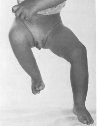
Figure 1.1 The Leg is short, flexed, abducted and externally rotated
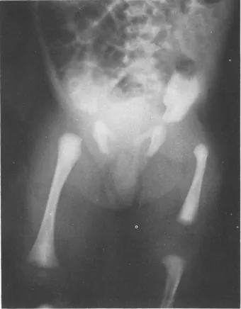
Figure 1.2 The proximal femur is translucent but the acetabulum is well formed. The femur has not moved proximally and its tip is bulbous being situated at a greater distance from the acetabulum than on the normal side
Associated abnormalities when present may be local or general. Tibial shortening with fibular dysplasia is more likely to be seen in company with either severe femoral deficiency or in association with simple short femur when it takes precedence and dominates the clinical problem (Hootnick, et al., 1976).
It would be surprising if an insult to the embryo during the stage of differentiation was localized in its effect and we must therefore be alert to the possibility of developmental anomalies elsewhere, especially in the arms.
Difficulty in diagnosis is unlikely when the manifestation is florid but in minor degrees, i.e. simple short femur and coxa vara, this is possible. A healed birth fracture will not show evidence of distal femoral epiphyseal delay and the shortening, if any, will not progress. Idiopathic hemiatrophy will declare itself in later childhood and is not identifiable in infancy, but the shortening and loss of girth characteristic of spinal dysraphism may be confusing as may unexplained dystrophy of a limb without the stigmata of femoral dysplasia. Congenital coxa vara must be distinguished from the infantile variety (see below).
CLASSIFICATION OF FEMORAL DYSPLASIA WITH SPECIAL REFERENCE TO THE PROGNOSIS OF THE HIP
It has already been emphasized that classification is essential to prognosis and prognosis is the key to management. Important contributions to the differentiation of types have been made by Aitken (1969) and Amstutz (1969) with others in a Symposium devoted to this subject which is probably the most significant publication on femoral dysplasia at the present time. Simple short femur and congenital coxa vara have been separately studied by Ring (1959, 1961) and Amstutz and Wilson (1962). The following observations are largely based upon the contributions of these authors.
Aitken’s classification distinguishes four subdivisions of established deformity of increasing severity with instability (A–D) (Figure 1.3). Amstutz provides this in more detailed form, also arranged in terms of progressively severe dysplasia, as Types I–V with subdivisions illustrating the initial state and the final outcome in all, with alternatives in two of the Types described (Figure 1.4). Although more complex, the Amstutz classification is more apposite to the practical problems of management especially as applied to the hip and will therefore be adopted in the following di...
Table of contents
- Cover image
- Title page
- Table of Contents
- POSTGRADUATE ORTHOPAEDICS SERIES
- Copyright
- Editor’s Foreword
- Chapter 1: Congenital Femoral Deficiency
- Chapter 2: Congenital Dislocation of the Hip and Associated Conditions
- Chapter 3: Pyogenic Arthritis of the Hip
- Chapter 4: Perthes’ Disease–Part I: Pathogenesis, Classification and Treatment
- Chapter 4: Perthes’ Disease–Part II: The Long-Term Results
- Chapter 5: Fractures of the Neck of the Femur in Children
- Chapter 6: Slipped Upper Femoral Epiphysis
- Chapter 7: Miscellaneous
- Index
Frequently asked questions
Yes, you can cancel anytime from the Subscription tab in your account settings on the Perlego website. Your subscription will stay active until the end of your current billing period. Learn how to cancel your subscription
No, books cannot be downloaded as external files, such as PDFs, for use outside of Perlego. However, you can download books within the Perlego app for offline reading on mobile or tablet. Learn how to download books offline
We are an online textbook subscription service, where you can get access to an entire online library for less than the price of a single book per month. With over 1.5 million books across 990+ topics, we’ve got you covered! Learn about our mission
Look out for the read-aloud symbol on your next book to see if you can listen to it. The read-aloud tool reads text aloud for you, highlighting the text as it is being read. You can pause it, speed it up and slow it down. Learn more about Read Aloud
Yes! You can use the Perlego app on both iOS and Android devices to read anytime, anywhere — even offline. Perfect for commutes or when you’re on the go.
Please note we cannot support devices running on iOS 13 and Android 7 or earlier. Learn more about using the app
Please note we cannot support devices running on iOS 13 and Android 7 or earlier. Learn more about using the app
Yes, you can access Hip Disorders in Children by G.C. Lloyd-Roberts,A.H.C. Ratliff, A. Graham Apley in PDF and/or ePUB format, as well as other popular books in Medicine & Orthopedics. We have over 1.5 million books available in our catalogue for you to explore.