- 368 pages
- English
- ePUB (mobile friendly)
- Available on iOS & Android
eBook - ePub
About this book
Forensic Dental Evidence: An Investigators Handbook highlights the discussion regarding unjust convictions caused by inaccurate bitemark opinions. The book focuses on cases that use forensic techniques, emphasizing modern methods and protocols. Through this book, the latest information available is offered to the forensic community.
This book demonstrates expertise in forensic dentistry by presenting chapters on human identification in domestic and international situations; investigations on missing person and violent crimes against persons; mass-disaster planning and disaster response; and new threats from terrorist attacks on urban centers. Furthermore, it discusses topics regarding bitemark evidence, such as forensic photography, analysis and legal issues. The book also presents two chapters on new scientific topics: The Next Level in Victim Identification: Materials Properties as an Aid in Victim Identification; and DNA for First Responders: Recognizing, Collecting, and Analyzing Biological Evidence Related to Dentistry (chapters 3 and 8, respectively).
This book is suited to anyone seeking knowledge on forensic dentistry; it will be of great value to investigators, lawyers, medical examiners, nurses, and dentists with an interest in forensic dental cases.
- Contributions by internationally recognized and experienced forensic experts cover missing persons cases and mass disaster cases from around the world
- Contains over 200 full-color photographs of crime scene evidence, human identification cases and bitemark details
- Includes many new exoneration cases derived from the Editor's work with the Innocence Project
Trusted by 375,005 students
Access to over 1.5 million titles for a fair monthly price.
Study more efficiently using our study tools.
Information
Chapter 1 Historical Dental Investigations
Dental Aging Analysis of Ancient Human Remains: Herakleides from the First Century AD
Overview
A death investigation of unidentified human remains requires professional determination of all physical evidence available from the body. The unknown person’s gender, age, medical status and cause of death are vital information for a forensic autopsy report. The following case explains details of the life and death of a young man whose body was originally found in Egypt. Dental information was important to confirm his age and give insight to his health history at the time of his death.
In 2003, the J. Paul Getty Museum Antiquities Conservation Department initiated the study of a Romano-Egyptian red-shroud mummy (91.AP.6) in the museum’s collection. The mummy is known as Herakleides from a painted inscription on top of the wrapped feet. Dating to the first century AD, the mummy incorporates a portrait panel depicting a young man in his early twenties. The mummy, measuring 175 cm in length, was wrapped in one large outer shroud that had been painted red and decorated with Egyptian funerary images. The beautifully executed portrait, the quality of the wrappings, and the elaborate use of gold, on both the panel and the shroud, attest to the prominent status of this individual.
The investigation of Herakleides’ mummy began with the conservation treatment of the fragile foot area, which was damaged and unstable [2]. The conservation treatment was in preparation for the mummy’s first public display at the Getty Villa in 2005, where its presence in the gallery contextualizes the now detached Romano-Egyptian portraits in the collection by illustrating their mortuary function. From here a full study of the body (human remains) and the materials used for the mummification and decoration of Herakleides evolved. The aim of this study was to better understand the person within the wrappings and the ancient techniques employed in its fabrication and adornment process. Imaging technology such as computerized tomography (CT) and infrared photographic techniques revealed secrets such as his complete name and the curious inclusion of a mummified ibis within the wrappings. Examination of the skeleton by orthopedic surgeons and a forensic dentist established his age, health, and height at the time of death. Radiocarbon dating (carbon 14) provided a secure date for the materials used in the mummification process and a likely time frame for when he lived. Motifs and religious iconography were studied and documented to better understand their meaning. The lack of clothing on the youth’s shoulders suggests he was an ephebe, or adolescent male of social standing. His presumed nudity, a symbol of rebirth, indicates he may have been an initiate in the cult of the Egyptian goddess Isis.
This study also involved the Getty Conservation Institute (GCI), which scientifically identified and compared the red pigment used on seven of the nine mummies identified within the red-shroud subgroup. The results from the analyses revealed that the composition of the unique red pigment is identical, relating this group to one another even further. The study of Herakleides shows how the collaboration of experts within the medical, scientific, and Egyptological communities can come together to better understand one unique artifact. This supportive exchange of experience, knowledge, and information has opened a window into the life, religion, and ritual of a man who lived almost 2,000 years ago [3].
The Forensic Examination of Herakleides
CT scans of the Herakleides’ mummy revealed that, contrary to the usual Egyptian practices of mummification, the 20-year-old man’s heart, not his lungs, were removed during embalming. Also uncommon in the scientists’ findings was a mummified ibis, inexplicably placed on Herakleides’ abdomen under the final layer of his mummy’s wrappings.
The Aging of Herakleides
Skeletal Analysis
His age determination was made by examining the epiphyses of his arms and legs. These are “growth plates” seen during teenage and early adult years that gradually disappear at maturity. They were faint in the Herakleides’ CT scan but were not completely fused. This is the data that produced an age range of 20 +/– 2 years. This opinion was provided by a radiologist at UCLA, who performed the CT scans, and was corroborated by two orthopaedic surgeons who examined Herakleides’ skeleton. There was no evidence of medical pathology (disease) or before-death (antemortem) trauma. A large gash is visible at the back of the skull, but it not clear whether this occurred before (antemortem) or after death (postmortem). The medical team who examined the CT scans believe it was most likely caused during mummification.
Dental Aging
Over the years, development of third molars (wisdom teeth) in adults has been researched in multiple population studies [4]. These studies compare the third molar root growth stages and development in the jawbone to the chronological (real age) of the known people in the study sample. Herekleides’ teeth are completely developed (the roots are fully formed) and are at the completed Stage H of full growth [5]. This indicates that he was at least 18 or older when he died. This supports the previous orthopaedic/anthropological opinions of this skeletal age at death.
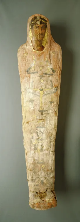
Figure 1.1 The mummy of Herakleides: “Mummy and Portrait on wooden panel.”
© The J. Paul Getty Museum, Villa Collection, Malibu, California (91.AP.6).
Acknowledgments
Photo: The J. Paul Getty Museum, Villa Collection, Malibu, California
References
[1] Corcoran L. Portrait Mummies from Roman Egypt (I-IV Centuries A.D.), with a catalogue of portrait mummies in Egyptian Museums. Chicago Press, 1995;7.
[2] . http://getty.edu/art/videos/mummification_process/mummification_process.html
[3] Corcoran L., Svoboda M. Herakleides: A Red-Shroud Portrait Mummy from Roman Egypt. Los Angeles: J. Paul Getty Museum, 2010.
[4] Bowers C.M. Determining Age from Teeth: The Estimation of Age From Dental Development. In: Bowers C.M., Bell G.L., editors. Manual of Forensic Odontology. third ed. ASFO; 1995:74-105.
[5] Ibid. p. 88.
The Odontological Identification of Adolf Hitler, Using Cinematographic Documents
Introduction
A “toothy” antemortem photograph can be invaluable when investigating the identity of unknown human remains. Pictures can show dental characteristics that can be very helpful to the forensic odontologist in comparing antemortem and postmortem data. Such documents can give a lot of information, but how much information is necessary for a positive identification? This paper gives investigators answers to this question from casework that combines both the photographic and dental evidence that is necessary for a positive determination of identity.
Forty-eight members of the sect of the Solar Temple were found dead in two different villages in October 1993. Their guru, Luc Jouret, was among them and was severely cremated. He was odontologically identified, and a picture published by the press was an additional contribution to the identification procedure [1].
The following account is a good example of how photographic documents may contribute to postulate an identity [2]. In October 2001, a Crossair plane crashed near the airport in Zurich, Switzerland. Eleven of the 24 passengers died in this mass disaster. The examination of the dental work of one passenger showed ceramic restorations that appeared unusual both in shape and shade. Before receiving any postmortem data of this otherwise cremated passenger, investigators were struck by an aspect of the dental work that led to the possibility tha...
Table of contents
- Cover Image
- Title page
- Copyright
- Dedication
- Contributing Authors
- Photo Credits
- Foreword
- Preface to Second Edition
- Preface to First Edition
- Acknowledgments
- Introduction
- Companion_website
- Table of Contents
- Chapter 1: Historical Dental Investigations
- Chapter 2: Dental Detectives
- Chapter 3: The Next Level in Victim Identification: Materials Properties as an Aid in Victim Identification
- Chapter 4: Forensic Dentistry Investigation Protocols
- Chapter 5: Recognition, Documentation, Evidence Collection, and Interpretation of Bitemark Evidence
- Chapter 6: Bitemarks in England and Wales
- Chapter 7: Legal Issues Concerning Bitemark Evidence in the United States
- Chapter 8: DNA for First Responders: Recognizing, Collecting, and Analyzing Biological Evidence Related to Dentistry
- Chapter 9: Missing and Unidentified Persons: The National Crime Information Center Dental Enhancements
- Chapter 10: The Disaster Victim Identification System: Its General Structure and the Swiss Involvement
- Chapter 11: Recognizing, Documenting, and Analyzing Physical Evidence in Abuse Cases
- Chapter 12: Managing a Mass Fatality Incident
- Chapter 13: Identifying Victims of 9/11: At the Office of Chief Medical Examine City of New York
- Chapter 14: Australasian and Multinational Disaster Victim Identification
- Chapter 15: Photography and Forensic Dental Evidence
- Chapter 16: The Use of Digital Imaging in Human Identification and Crime Scene Analysis
- Index
- Dental Investigations in Mass Disaster Incidents
- Violence Against Persons: Images and Terminology for Investigators
- Atlas of Dental Identification
Frequently asked questions
Yes, you can cancel anytime from the Subscription tab in your account settings on the Perlego website. Your subscription will stay active until the end of your current billing period. Learn how to cancel your subscription
No, books cannot be downloaded as external files, such as PDFs, for use outside of Perlego. However, you can download books within the Perlego app for offline reading on mobile or tablet. Learn how to download books offline
Perlego offers two plans: Essential and Complete
- Essential is ideal for learners and professionals who enjoy exploring a wide range of subjects. Access the Essential Library with 800,000+ trusted titles and best-sellers across business, personal growth, and the humanities. Includes unlimited reading time and Standard Read Aloud voice.
- Complete: Perfect for advanced learners and researchers needing full, unrestricted access. Unlock 1.5M+ books across hundreds of subjects, including academic and specialized titles. The Complete Plan also includes advanced features like Premium Read Aloud and Research Assistant.
We are an online textbook subscription service, where you can get access to an entire online library for less than the price of a single book per month. With over 1.5 million books across 990+ topics, we’ve got you covered! Learn about our mission
Look out for the read-aloud symbol on your next book to see if you can listen to it. The read-aloud tool reads text aloud for you, highlighting the text as it is being read. You can pause it, speed it up and slow it down. Learn more about Read Aloud
Yes! You can use the Perlego app on both iOS and Android devices to read anytime, anywhere — even offline. Perfect for commutes or when you’re on the go.
Please note we cannot support devices running on iOS 13 and Android 7 or earlier. Learn more about using the app
Please note we cannot support devices running on iOS 13 and Android 7 or earlier. Learn more about using the app
Yes, you can access Forensic Dental Evidence by C. Michael Bowers in PDF and/or ePUB format, as well as other popular books in Law & Forensic Science. We have over 1.5 million books available in our catalogue for you to explore.