eBook - ePub
Structural Biology in Immunology
Structure/Function of Novel Molecules of Immunologic Importance
- 186 pages
- English
- ePUB (mobile friendly)
- Available on iOS & Android
eBook - ePub
Structural Biology in Immunology
Structure/Function of Novel Molecules of Immunologic Importance
About this book
Structural Biology in Immunology, Structure/Function of Novel Molecules of Immunologic Importance delivers important information on the structure and functional relationships in novel molecules of immunologic interest. Due to an increasingly sophisticated understanding of the immune system, the approach to the treatment of many immune-mediated diseases, including multiple sclerosis, systemic lupus erythematosus, rheumatoid arthritis, and inflammatory bowel disease has been dramatically altered. Furthermore, there is an increasing awareness of the critical role of the immune system in cancer biology. The improved central structure function relationships presented in this book will further enhance our ability to understand what defects in normal individuals can lead to disease.
- Describes novel/recently discovered immunomodulatory proteins, including antibodies and co-stimulatory or co-inhibitory molecules
- Emphasizes new biologic and small molecule drug design through the exploration of structure-function relationship
- Features a collaborative editorial effort, involving clinical immunologists and structural biologists
- Provides useful and practical insights on developing the necessary links between basic science and clinical therapy in immunology
- Gives interested parties a bridge to learn about computer modeling and structure based design principles
Trusted by 375,005 students
Access to over 1.5 million titles for a fair monthly price.
Study more efficiently using our study tools.
Information
Topic
MedicineSubtopic
ImmunologyChapter 1
Organization of Immunological Synapses and Kinapses
Marco Fritzsche*,†; Michael L. Dustin*,‡ * Kennedy Institute of Rheumatology, Oxford, United Kingdom,
† Weatherall Institute of Molecular Medicine, University of Oxford, Oxford, United Kingdom,
‡ New York University School of Medicine, New York, NY, United States
† Weatherall Institute of Molecular Medicine, University of Oxford, Oxford, United Kingdom,
‡ New York University School of Medicine, New York, NY, United States
Abstract
Immunological synapses and kinapses describe two modes through which cells of the immune system exchange information based on specific recognition. This chapter focuses on T-cell immunological synapses, particularly, the structural aspects building from molecules that undergo stepwise assembly into the complex supramolecular activation clusters that captivated biology 20 years ago. Modern imaging methods have provided insights into the linkage between physical processes such as liquid-phase separation in adhesion, membrane, and signaling layers of the interface. These processes then link to the F-actin cortex and dramatic changes in membrane topology emerging from exocytic, endocytic, and extracellular vesicle budding mechanisms to shape and maintain this intricate communication interface.
Keywords
Synapse; Kinapse; Signaling; Activation; Phase separation; Actin; Cortex; Extracellular vesicles; Exosomes; Ectosomes; Exocytosis
Abbreviations
Ag antigen
Arp actin-related protein
CD(#) cluster of differentiation (1,2,3…)
CRTAM class-I MHC-restricted T-cell-associated molecule
CSK C-terminal Src kinase
CTLA-4 cytotoxic T lymphocyte antigen-4
DC dendritic cell
ICAM(#) intercellular adhesion molecule-(1,2,3…)
ICOS(L) inducible T-cell costimulator (ligand)
ITAM immunotyrosine activation motif
LAT linker of activated T cells
LFA-(#) lymphocyte function associated-(1,2,3)
(p)MHC (peptide) major histocompatibility complex
PD-1 programmed cell death-1
SH(#) Src homology domain (1,2,3)
SIM structured illumination microscopy
SLAM signaling lymphocyte activation molecule
SLB supported lipid bilayer
(c,p,d)SMAC (central, peripheral, distal) supramolecular activation cluster
STED stimulated excitation depletion
TCR T-cell antigen receptor
(e)TIRF(M) (extended) total internal reflection fluorescence (microscopy)
VASP vasodilator-stimulated phosphoprotein
WASp Wiscott Aldrich syndrome protein
ZAP-70 TCR zeta-associated protein of 70 kili-Daltons
Acknowledgments
We thank our groups for stimulating data and discussions. MLD was supported by Wellcome Trust and Kennedy Trust for Rheumatology Research (PRF 100262Z/12/Z).
1.1 Introduction
Immunological synapses are stable adhesive junctions formed by immune cells for priming of immune responses and effector function.1 The archetype is based on radially symmetric junctions formed by helper or cytotoxic T cells, with B cells as antigen-presenting cells in which concentric zones referred to as supramolecular activation clusters (SMACs) can be resolved by wide-field microscopy in vitro and in vivo.2,3 The most important function of the immunological synapses is directed secretion of regulatory signals in the form of proteins and vesicles between immune cells, and also killing of pathogens in the context of innate immune recognition (,4–7 p. 5140). Immunological synapses are formed by T cells, innate lymphoid cells, B cells, mast cells, neutrophils, macrophages (phagocytic synapse), and likely others.5,7–10 The key common denominator is that specificity is driven by receptors that couple to nonreceptor tyrosine kinases, often through immunotyrosine activation motifs in the cytoplasmic domain.11 The T-cell antigen receptor (TCR) is the relevant receptor for T cells,12 which will be the major focus of this chapter.
The formation of the archetypal T-cell immunological synapse can also be reconstituted with supported lipid bilayers (SLBs) containing major histocompatibility complex (MHC) proteins with bound agonist peptides (pAgMHC) and the adhesion molecule intercellular adhesion molecule-1 (ICAM-1, CD54) in which SMACs form in a time-dependent manner.10 The two major SMACs are the central SMAC (cSMAC), which is enriched in T-cell receptors (TCRs), pAgMHC, and some costimulatory receptors such as CD28, and the peripheral SMAC (pSMAC) that is enriched in the integrin lymphocyte function associated 1 (LFA-1, CD11a) and ICAM-12 (Fig. 1.1A). Subsequently, Freiberg et al. defined the distal SMAC (dSMAC) as a ring outside the pSMAC enriched in CD45 and dynamic filamentous actin (F-actin).13,14 Some aspects of immunological synapse formation can also be reconstituted with glass substrates coated with anti-CD3 antibodies to trigger TCR signals and other surface treatments, such as poly-l-lysine or serum attachment factors (fibronectin, vitronectin, etc.) that provide an adhesion component.15,16 In the absence of this adhesion component, anti-CD3 elicits actin-rich protrusions that push the cell away from the anti-CD3-coated surface.17,18 We will incorporate data from cell-cell systems and model substrates to generate a current picture and trajectory for future questions.
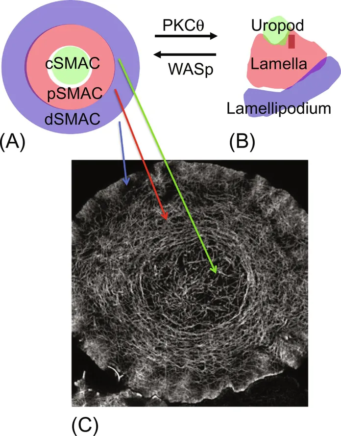
Stability of the immunological synapse depends on its symmetry. Breaking of the symmetry of the immunological synapse leads to a migratory junction defined as a kinapse, which appears to be an important mode of T-cell interaction with, for example, dendritic cells (DCs) and germinal center B cells in vivo10,13,19–23 (Fig. 1.1B). Kinapse formation may lead to “multifocal synapses” observed in T-DC interactions in vitro and in vivo.24,25 The immunological synapse and kinapse share signaling elements, referred to as microclusters, which are formed by both the TCR and LFA-1.26–29 While the formation of microclusters likely has some aspects driven by the biophysics of receptor- ligand-driven self-assembly in the interface, most aspects of immunological synapse and kinapse formation and maturation are dependent upon intact F-actin cytoskeleton and later stages of synapse maturation are regulated by microtubules.30–32 The chapter will build a structural model for the immunological synapse beginning with some major classes of receptors and then moving to the underlying cytoskeletal networks and important issues of membrane topology.
1.2 Receptors and Ligands of the Immunological Synapse and Kinapse
1.2.1 T-cell Receptor and pAgMHC
There are two known TCR heterodimers—αβ and γδ—that are expressed on distinct subsets of T cells.12 The classical adaptive T-cell subsets use the αβ TCR, whereas subsets of tissue-resident T cells use the γδ TCR.33 Most γδ T cells and a small subset of αβ T cells are innate like, meaning that they use stereotypical rearrangements to generate largely invariant receptors for conserved ligands that are often presented on nonclassical MHC-like proteins. For example, the so-called natural killer T cells use invariant αβ TCR to recognize glycolipids bound to CD1d, which has a structure similar to MHC class I, but possesses a binding groove adapted for the recognition of glycolipids.34 Mucosal-associated invariant T cells recognize microbial metabolites presented on MHC-related protein.35 This chapter focuses on the majority of αβ T cells that use MHC class I or class II proteins as ligands in combinations with short peptides. Each of these αβ T cells expresses one TCR that binds weakly to self-peptide (pself)-MHC based on thymic selection and has the potential to bind a variety of pAgMHC.36 Since each TCR is generated through multiple recombination events involving germline V, (D), and J segments with random addition of base pairs during joining, there is huge diversity of these weakly self-reactive TCR in the “naïve” repertoire that are selected for expansion and conversion into memory T cells over the life of an individual. The interaction of TCR with pself–MHC is challenging to measure physically using the available methods, but in solution have dissociation constants on the order of 100 μM to > 1 mM,37,38 yet these interactions are essential for thymic selection during development and appear to influence mature T cells continuously.39 The interactions of TCR with pAgMHC typically have Kd in the range of 100 nM–10 μM.38 All individuals seem to harbor some higher affinity pself-MHC-specific T cells that often take on suppressive roles as regulatory T cells (Treg),40 but can also be rec...
Table of contents
- Cover image
- Title page
- Table of Contents
- Copyright
- Dedication
- Contributors
- Acknowledgments
- Chapter 1: Organization of Immunological Synapses and Kinapses
- Chapter 2: Principles of Protein Recognition by Small T-Cell Adhesion Proteins and Costimulatory Receptors
- Chapter 3: Synthetic Antibody Engineering: Concepts and Applications
- Chapter 4: Natural Killer Cell Receptors
- Chapter 5: Structure-Function in Antibodies to Double-Stranded DNA
- Chapter 6: The Role of the Constant Region in Antibody-Antigen Interactions: Redefining the Modular Model of Immunoglobulin Structure
- Index
Frequently asked questions
Yes, you can cancel anytime from the Subscription tab in your account settings on the Perlego website. Your subscription will stay active until the end of your current billing period. Learn how to cancel your subscription
No, books cannot be downloaded as external files, such as PDFs, for use outside of Perlego. However, you can download books within the Perlego app for offline reading on mobile or tablet. Learn how to download books offline
Perlego offers two plans: Essential and Complete
- Essential is ideal for learners and professionals who enjoy exploring a wide range of subjects. Access the Essential Library with 800,000+ trusted titles and best-sellers across business, personal growth, and the humanities. Includes unlimited reading time and Standard Read Aloud voice.
- Complete: Perfect for advanced learners and researchers needing full, unrestricted access. Unlock 1.5M+ books across hundreds of subjects, including academic and specialized titles. The Complete Plan also includes advanced features like Premium Read Aloud and Research Assistant.
We are an online textbook subscription service, where you can get access to an entire online library for less than the price of a single book per month. With over 1.5 million books across 990+ topics, we’ve got you covered! Learn about our mission
Look out for the read-aloud symbol on your next book to see if you can listen to it. The read-aloud tool reads text aloud for you, highlighting the text as it is being read. You can pause it, speed it up and slow it down. Learn more about Read Aloud
Yes! You can use the Perlego app on both iOS and Android devices to read anytime, anywhere — even offline. Perfect for commutes or when you’re on the go.
Please note we cannot support devices running on iOS 13 and Android 7 or earlier. Learn more about using the app
Please note we cannot support devices running on iOS 13 and Android 7 or earlier. Learn more about using the app
Yes, you can access Structural Biology in Immunology by Chaim Putterman,David Cowburn,Steven Almo in PDF and/or ePUB format, as well as other popular books in Medicine & Immunology. We have over 1.5 million books available in our catalogue for you to explore.