![]()
CHAPTER 1
Functional Anatomy of the Myocardium
1. Preliminary considerations
Classical anatomy of the heart considered that the muscle structure forming the myocardium was homogeneous and compact. Based on this concept, it was described by an external and internal surface limiting a solid, uniform muscle mass. Andreas Vesalius, in his work De Humanis Corporis Fabrica (1543), referred to the difficulty in identifying the layers forming the myocardium. He literally expressed “Whichever way you perform a dissection of the heart meat, either raw or cooked…, you can hardly remove a portion with only one type of fiber, because they have multiple and different directions, mainly transversal”. Three centuries after (1864), J. B. Pettigrew also referred to this situation: “About the complexity of the arrangement I need not say more than Vesalius, Haller and De Blainville, who all confessed their inability to elucidate it”. (81, 114)
This classical structural notion does not explain cardiac mechanics; hence, it is essential to establish its true internal anatomy. Historically, very little importance was attributed to the spatial arrangement of the muscle bundles forming the myocardium. (23) In1970 Francisco Torrent Guasp (102-108) defines the anatomy of the heart adapted to physiological reality. This study approach correlates with a cardiac structure presenting the remarkable characteristic of being adriving-suction pump with the size equivalent to a human fist and an average weight of 270 grams, which ejects 4-6 liters/minute at a speed of 300 cm/s, consuming only 10 watts, and working without interruption for 80 years without maintenance, almost without noise, and no smoke. Its work is equivalent to the daily withdrawal of 1 ton of water 1 m deep with a mechanical efficiency (work/energy relationship) of 50%, not achieved by man-made machines which only attain 30% efficacy. This allows ejecting 70% of the left ventricular volume with only 12% shortening of its contractile unit, the sarcomere.
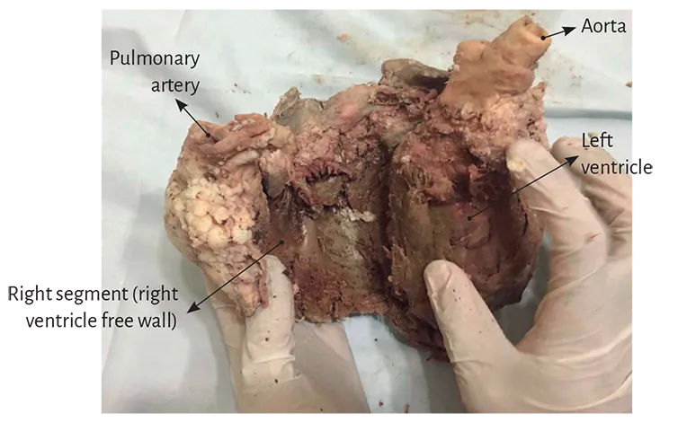
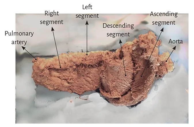
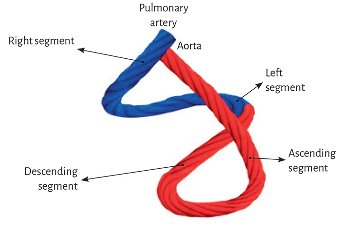
Torrent Guasp demonstrated through numerous dissections of hearts from different species, including humans, that the ventricular myocardium is formed by an assembly of fibers coiled unto themselves similarly to a rope (rope model) (Figures 1.1 to 1.4) and flattened laterally as a band, which by giving two spiral turns defines a helix limiting both ventricles and setting their performance. (2, 3) This structure is supported by the evolutionary process taking place from the primitive circulatory duct of the annelids to that of mammals, whose arterial circuit forms a loop or fold that twists unto itself giving rise to the ventricular chambers. The primary duct lumen establishes a secondary communication between both adjacent chambers (ventricles) formed by the loop, assuming that the side of the duct where the interconnection takes place must have cleaved all along the duct to achieve this purpose.
Based on these facts, we find that the spatial organization and the rotational motion of the ventricular fibers in their anatomical arrangement, both at the base and apical region levels, find correspondence with the myocardial band. However, after its original description, this anatomy which allows unfolding the heart to form a muscle band has not been considered valid until now by the medical culture.
Figure 1.1. Myocardial band.
Figure 1.2. Origin of myocardial band unfolding.
Figure 1.3. Myocardial band prior to its
complete unfolding (See Figure 1.12).
Figure 1.4. Rope model of the myocardial band.
It illustrates the different segments that form the band.
In blue: Basal loop. In red: Apical loop.
In view of the criticism or indifference met by the helical myocardial band proposed by Torrent Guasp due to lack of information on the necessary anatomical technique to unfold it, we can currently obtain its confirmation through:
- Anatomical and histological study of the heart. (128)
- The evolutionary concept emerging from phylogeny.
- New imaging procedures obtained with magnetic resonance imaging by diffusion tensor. (17, 24, 82, 142)
- Echocardiography. (59, 70, 71)
- Electrophysiological studies performed with three-dimensional electroanatomical mapping. (121-127, 131, 132)
Regarding the argued difficulty to dissect the myocardium, which is more apparent than real (128), we should consider that once the myocardial band originated as a loop in the arterial semicircle of amphibians and reptiles to adapt to the physiological demands of aerial life, the muscle bundles became firmly attached to their contact surfaces, hampering the necessary cleavage planes necessary for their anatomical dissection. The evolutionary goal was to develop a sufficiently solid hemodynamic structure with the strength to generate the suction and pumping of the blood volume that supplied the whole organism. Thus, every attempt to dissect an anatomical segment from the rest of the myocardium, avoiding the real cardiac arrangement, always turns into an obstacle due to the structural plan of the axes where the orientation of the myocardial band courses.
An explanation for this muscular homogeneity hiding the myocardial band, implies considering the required functioning in birds and mammals to achieve blood ejection at a high speed in a limited time span by an organ that must supply two circulations (systemic and pulmonary). Despite these considerations and the classical concept about myocardial anatomy, its dissection finds a structure with defined planes where the successive and related physiological heart motions of narrowing, shortening-twisting, lengthening-untwisting and expansion take place depending on the propagation of the electrical stimulus along its muscle pathways (chapter 1). (125)
The myocardial fibers forming the myocardium cannot be considered as absolutely independent entities within a defined space. Despite the intricacy of fiber bundles with polygonal shape, which in addition receive and give off collateral fibers, a predominant course of central fibers is defined with sliding planes, which together form the myocardial muscle band. It should be recalled that the myocardium constitutes a spiraling continuum in its fibers responding to the helical pattern in its muscle bundles. This arrangement indicates the need of generating a mechanical work that dissipates little energy. Therefore, the fiber layers very gradually shift their orientation, with more or less acute angles, to avoid that abrupt changes in the spatial organization dissipate the necessary work for cardiac function. The fan of fibers that is formed reduces the stress among them.
This situation generates a tangle of fibers that allows the band to behave as a continuous transmission chain with the epicardial fibers taking an oblique direction, the intermediate fibers a transverse course and the endocardial fibers also an oblique direction, but contrary to that of the epicardial plane. The endocardial and epicardial plane access angle is approximately 60 degrees in relation to the transverse fibers. Fiber orientation defines function and thus the ejection fraction is 60% when the normal helical fibers contract and falls to nearly 30% if only the transverse fibers shorten. This occurs when the left ventricle dilates in cardiac remodeling and the fibers miss their oblique orientation, loosing muscular and mechanical efficiency.
It should therefore be acknowledged that a gradual change in orientation is generated from the superficial to the deep fibers that form the different segments of the muscle band. In the progression from the ventricular base to the apex, the number of horizontal fibers decreases in relation to the oblique fibers, showing that the heart is organized as a continuous muscle helix. The ventricular mechanical activity must be heterogeneous during diastole with subendocardial-subepicardial relaxation gradients. During systole, the muscle layers of the myocardial band evidence pronounced and opposite torsion in the subendocardium in relation to the subepicardium, whereas in the apex the subepicardial fiber rotation acquires more relevance.
Beyond this complexity it is necessary to establish the concept of linear and laminar trajectories. Myocardial muscle bundles and bands, which derive from phylogenetic development, essentially shape a master axis of precise dynamic requirement. The spatial muscle structure adopted by the myocardial muscle band has a double function: a) to limit the ventricular chambers and b) to fulfill the suction and driving action in its role of cardiac pump.
2. Myocardial architecture
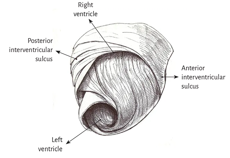
Left ventricle. The entire apex belongs to the left ventricle. In the distal part of the left ventricle, called apical, a muscle layer with spiral trajectory extends from the surface to the center and undergoes a rotation that turns the subepicardial fibers into subendocardial fibers, overlapping like the tiles of a roof. Consequently, the left ventricular distal end, the apex, surrounds a virtual conduit with no muscular plane at its ultimate end, lined internally by the endocardium and externally by the epicardium, with no intermediary muscle. It is essential to consider that in the apex the fibers undergo a helical rotational motion with sphincter-like arrangement as they transform from subepicardial to subendocardial fibers, following a clockwise trajectory (apical view of the diaphragmatic surface of the heart in the anatomical position) (Figures 1.5 and 1.6). (105)
Figure 1.5. Apical view of the left and right ventricles.
Figure 1.6. Spiral arrangement of apical muscle layers.
In the left ventricular basal half, at the level of its free wall (Figure 1.7), the fibers are arranged similarly to the apical half. A muscle layer with spiral trajectory extends from the surface to the center displaying its fibers from outside to inside (from paraepicardial to paraendocardial regions). At this level their orientation is opposite to that of the apex, following a counter-clockwise trajector...