The Pocket Guide to Diagnostic Cardiac Catheterization provides general cardiology fellows, nurses, and technicians entering the cardiac catheterization laboratory a practical guide addressing key aspects of left and right heart catheterization, selective coronary angiography, and the utilization of other invasive cardiology procedures for diagnostic purposes.Written by a team of physicians from Emory University Hospital's Cardiac Catheterization Laboratories, practical pearls of wisdom about the technical aspects of cardiac catheterization and other invasive cardiology procedures are presented with step-by-step instructions and easy-to-follow illustrations in this quick reference of essential material.Also included is a chapter with video clips that highlight the role and technical versatility of the multipurpose catheter in cardiac catheterization—a technique developed, taught, and learned over many generations in the cardiac catheterization laboratories at Emory University.
eBook - ePub
Pocket Guide to Diagnostic Cardiac Catheterization
- 306 pages
- English
- ePUB (mobile friendly)
- Available on iOS & Android
eBook - ePub
Pocket Guide to Diagnostic Cardiac Catheterization
About this book
Trusted by 375,005 students
Access to over 1.5 million titles for a fair monthly price.
Study more efficiently using our study tools.
Information
Topic
MedicineSubtopic
CardiologyCHAPTER 1
A Brief History of Cardiac Catheterization
“If you want to understand anything, observe its beginning and its development.”
—Aristotle
History of a Procedure
A reader who flips through the first pages of this Pocket Guide might ask, “Do I really have to read this chapter to learn how to perform cardiac catheterization?” The rational answer is, “No.” Nonetheless, please be irrational; do not skip these pages. You will be able to review the fascinating story of human curiosity, courage, and dedication of physicians and scientists who pursued their work despite the prevailing clinical and scientific dogmas to set the stage for a new generation of investigators who moved the field from diagnostic to therapeutic procedures.
No one can accurately pinpoint the exact time when humans started to be interested in cannulating blood vessels and cardiac chambers, but it is known that ancient Egyptians, Greeks, and Romans were forming tubes from hollow reeds, palm leaves, and pipes to study the function of cardiac valves in cadavers.1 Many centuries later in 1733, British physiologist Stephen Hales performed the first catheterization of the arterial blood vessel in a horse using brass pipes and a glass tube.2 In 1844, French physiologist Claude Bernard was first to catheterize the left and right ventricles in animals by inserting a mercury thermometer into the carotid artery and jugular vein.3 Adolph Fick came up with a brilliant one-page note on the calculation of blood flow in 1870, which opened the experimental era of cardiac metabolism.4 Finally, in Eberswalde, Germany in 1929, Werner Forssmann self-cannulated his antecubital vein and guided a urological catheter into his right atrium documenting its location via chest x-ray.5 In 1941, Andre Cournand and Dickinson Richards began to utilize right heart catheterization to study cardiac output, and in 1956 they shared the Nobel Prize in Physiology and Medicine with Forssmann.
Lewis Dexter discovered in 1948 that by wedging the catheter into a distal branch of a pulmonary artery, it is feasible to record the height of the left atrial pressure.6 In the late 1960s, Jeremy Swan and William Ganz used a balloon-tipped flow-directed catheter in the right heart to continuously measure and monitor hemodynamic tracings.7 Ganz improved the catheter design by adding the ability to measure cardiac output using the thermodilution method. Henry Zimmerman, working at the Cleveland Clinic, is credited with combined right and left heart catheterization in 1947.8 In 1952, Sven-Ivar Seldinger from Sweden came up with a simple and brilliant idea of over the wire insertion of a catheter, which revolutionized the approach for cannulation of arteries and veins.9 In 1958, Mason Sones at the Cleveland Clinic developed a selective technique to cannulate coronary arteries.10 In 1966, Melvin Judkins introduced his method for transfemoral selective coronary arteriography and subsequently popularized his first pre-formed left and right coronary catheters to simplify the process of selective cannulation of each coronary ostium.11 Kurt Amplatz developed his own pre-formed coronary catheters in 1967.12 In the late 1960s, Fred Schoonmaker and Spencer King introduced a single-catheter technique with a modified Sones-type catheter and published results of its use in 1974.13
In 1964, Charles Dotter and Melvin Judkins treated obstructed atherosclerotic peripheral arteries by using coaxial catheters to dilate their lumen to the size of 12- to 14-French (Fr) catheters.14 In 1973, Werner Porstmann15 and Eberhard Zeitler16 independently published data of using a balloon technique to dilate a blood vessel, but it was Andreas Roland Gruentzig who successfully performed the first percutaneous coronary balloon angioplasty in 1977 in a 38-year-old man with an 85% stenosis in the mid segment of the left anterior descending coronary artery, thus opening the door to the fascinating era of interventional cardiology.17
References
1.Miller SW. Cardiac angiography. Boston, MA: Little, Brown; 1984.
2.Hales S. Statistical essays, containing haemastaticks. Vol 2. London: W Innys, R Manby, T Woodward; 1733.
3.Cournand AF. Cardiac catheterization: development of the technique its contributions to experimental medicine, and its initial applications in man. Acta Med Scand. 1975;579(suppl):7-32.
4.Fick A. Über die Messung des Blutquantums in den Herzventrikeln. Sitzungsber. Phys-Med Ges Würzburg; 1870.
5.Forssmann W. Die Sondierung des rechten Herzens. Klin Wschr. 1929;8:2085-2087.
6.Dexter L, Hayes FW, Burwell CS, Eppinger EC, Sagerson RP, Evans JM. Studies of congenital heart disease: II. The pressure and oxygen content of blood in the right auricle, right ventricle, and pulmonary artery in control patients, with observations on the oxygen saturation and source of pulmonary ‘capillary’ blood. J Clin Invest. 1947;26:554-560.
7.Swan HJC, Ganz W, Forrester J, Marcus H, Diamond G, Chonette D. Catheterization of the heart in man with use of a flow directed balloon-tipped catheter. N Engl J Med. 1970;283:447-451.
8.Zimmerman HA, Scott RW, Becker NO. Catheterization of the left side of the heart in man. Circulation. 1950;1:357-359.
9.Seldinger SI. Catheter replacement of the needle in percutaneous arteriography: a new technique. Acta Radiol. 1953;39:368-376.
10.Sones FM Jr., Shirey EK, Proudfit WL, Westcott RN. Cine-coronary arteriography (Abstract). Circulation. 1959;20:773.
11.Judkins MP. Selective coronary arteriography: a percutaneous transfemoral technique. Radiology. 1967;89:815-824.
12.Wilson WJ, Lee GB, Amplatz K. Biplane selective coronary arteriography via percutaneous transfemoral approach. Am J Roentgenol. 1967;100:332-340.
13.Schoonmaker FW, King SB. Coronary arteriography by the single catheter percutaneous femoral technique. Experience in 6800 cases. Circulation. 1974;50:735-740.
14.Dotter CT, Judkins MP. Transluminal treatment of arteriosclerotic obstruction: description of a new technique and a preliminary report of its application. Circulation. 1964;30:654-670.
15.Porstmann W. Ein neuer Korsett-Ballonkatheter zur transluminalen Rekanalisation nach Dotter unter besonderer Berücksichtigung von Obliterationen an den Beckenarterien. Radio Diagn. 1973;14:239-244.
16.Zeitler E. Percutaneous treatment of arterial blood circulation disorders of the extremities using a catheter. Radiologe. 1973;13(8):319-324.
17.Gruentzig A. Transluminal dilatation of coronary-artery stenosis [Letter]. Lancet. 1978;1:263.
CHAPTER 2
The Cardiac Catheterization Laboratory
“Any sufficiently advanced technology is indistinguishable from magic.”
—Arthur C. Clarke
Catheterization Laboratory Equipment
Most cardiac catheterization laboratories consist of two separate rooms. The control room is used for monitoring the patient’s vital signs, hemodynamics, and ECG tracings. Here, physicians discuss the planned procedure and write the final report. Next to the control room is the main laboratory (Figure 2.1).
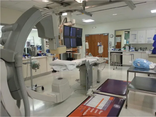
FIGURE 2.1A contemporary cardiac catheterization laboratory.
The examination table can be ground-mounted or suspended from the ceiling at one end; it is movable and can be panned, raised, or lowered. The table is centered between a C-shaped structure called a gantry, which houses the laboratory’s radiographic equipment. At the bottom of the gantry is the x-ray source, composed of both an x-ray generator and an x-ray tube. At the top of the gantry, or C-arm, there is an “eye-eye,” the flat detector that provides high-resolution digital imagery. Other equipment located throughout the lab may include, but is not limited to, the following important items: an intra-aortic balloon pump and a code or “crash” cart with a defibrillator. In the catheterization laboratory, invasive blood pressure is monitored, and pressure waves created by cardiac contractions are transmitted through a fluid-filled tube connected to a pressure transducer, which converts the actual pressure waveforms into electrical signals displayed on monitors. One other piece of equipment is the power injector, which allows the physician to administer contrast rapidly and in large volumes.
The catheterization laboratory team is composed of several key positions. First, the monitoring person who in the control room closely monitors the procedure, continuously documents vital signs, ECG, hemodynamic parameters of the patient, and informs the operator of any abnormalities noticed. This position is staffed by a nurse or by a catheterization laboratory/radiology technician who is knowledgeable in cardiac hemodynami...
Table of contents
- Cover
- Dedication
- Table of Contents
- About the Authors
- Foreword
- Preface
- Abbreviations
- Video Files of Multipurpose (MP) Catheter Manipulation
- The Laws of Dr. F. Mason Sones
- 1. A Brief History of Cardiac Catheterization
- 2. The Cardiac Catheterization Laboratory
- 3. The Tools
- 4. Precatheterization Care
- 5. Vascular Access
- 6. Coronary, Renal, and Mesenteric Angiography
- 7. The Multipurpose Catheter
- 8. Angiography of Coronary Bypass Grafts
- 9. Left and Right Ventriculography, Aortography, and Pulmonary Angiography
- 10. Right Heart Catheterization
- 11. Right and Left Heart Hemodynamics
- 12. Shunt Detection and Calculation
- 13. Endomyocardial Biopsy
- 14. Pericardiocentesis
- 15. Intra-Aortic Balloon Pump (IABP) Placement
- 16. Temporary Transvenous Pacemaker Placement
- 17. Post-Cardiac Catheterization Care
- 18. Approach to Complex Cases in Cardiac Catheterization
- 19. Useful Formulae and Normal Values
- Index
Frequently asked questions
Yes, you can cancel anytime from the Subscription tab in your account settings on the Perlego website. Your subscription will stay active until the end of your current billing period. Learn how to cancel your subscription
No, books cannot be downloaded as external files, such as PDFs, for use outside of Perlego. However, you can download books within the Perlego app for offline reading on mobile or tablet. Learn how to download books offline
Perlego offers two plans: Essential and Complete
- Essential is ideal for learners and professionals who enjoy exploring a wide range of subjects. Access the Essential Library with 800,000+ trusted titles and best-sellers across business, personal growth, and the humanities. Includes unlimited reading time and Standard Read Aloud voice.
- Complete: Perfect for advanced learners and researchers needing full, unrestricted access. Unlock 1.5M+ books across hundreds of subjects, including academic and specialized titles. The Complete Plan also includes advanced features like Premium Read Aloud and Research Assistant.
We are an online textbook subscription service, where you can get access to an entire online library for less than the price of a single book per month. With over 1.5 million books across 990+ topics, we’ve got you covered! Learn about our mission
Look out for the read-aloud symbol on your next book to see if you can listen to it. The read-aloud tool reads text aloud for you, highlighting the text as it is being read. You can pause it, speed it up and slow it down. Learn more about Read Aloud
Yes! You can use the Perlego app on both iOS and Android devices to read anytime, anywhere — even offline. Perfect for commutes or when you’re on the go.
Please note we cannot support devices running on iOS 13 and Android 7 or earlier. Learn more about using the app
Please note we cannot support devices running on iOS 13 and Android 7 or earlier. Learn more about using the app
Yes, you can access Pocket Guide to Diagnostic Cardiac Catheterization by Andro G. Kacharava,Stephen D. Clements,A. Maziar Zafari in PDF and/or ePUB format, as well as other popular books in Medicine & Cardiology. We have over 1.5 million books available in our catalogue for you to explore.