- 46 pages
- English
- ePUB (mobile friendly)
- Available on iOS & Android
eBook - ePub
Specimen Preparation for Transmission Electron Microscopy of Materials
About this book
Details the essential practical steps which must precede microscopy. Methods for preparing sheet or disc specimens and final thinning techniques are described with reference to practical problems. The book also covers methods for mounting specimens in the
Trusted by 375,005 students
Access to over 1.5 million titles for a fair monthly price.
Study more efficiently using our study tools.
Information
1
The specimen: what we are trying to achieve
Electron microscopy is a powerful and fascinating tool for the investigation of structure and composition on a fine scale. Considerable skill is required to operate the microscope itself, to take high quality micrographs, and to interpret the resultant images. These topics are dealt with in companion handbooks. In this volume we shall be concentrating on the essential practical steps which must precede the microscopy itself and which are absolutely crucial to successful and meaningful use of a transmission electron microscope. Without a thin undamaged specimen even the most skilful microscopist is helpless.
Let us first state the primary objective of specimen preparation. It is to prepare and mount successfully in the microscope a thin specimen from which it will be possible to deduce accurately the structure, composition, and often also the behaviour of a larger sample of the material. There are several possible general approaches to achieving this objective. The most common is to thin a large piece of the material, for example by electropolishing, or to disperse it into pieces which are small enough to be transparent to electrons. This is the subject of Chapters 2 and 3. However, two alternative approaches are useful in a limited number of cases. It is sometimes possible to prepare the material directly in the form of a thin sheet, for example by vacuum deposition. Indeed, this often produces a marvellous specimen in all respects except the most important — it may not be representative of the material on a larger scale. However, if the object is to study thin deposited films then this simple technique is clearly highly appropriate. A third approach to specimen preparation is to extract from a larger specimen only certain components of its structure for study in isolation. This can be convenient but of course cannot give a total picture of the microstructure. The major technique of this type is carbon extraction replication, which is dealt with in Chapter 4.
1.1 Thinking ahead
Before deciding on a specimen preparation route (that is, if it is not already too late, before going straight to Chapter 3) it is wise to consider what transmission electron microscopy (TEM) techniques are to be used on the specimen. This will help to decide what are going to be the most important attributes of the final foil. For example, if large-scale low magnification information about the structure is required, uniformity of thinning and large size of thin area are paramount. On the other hand for weak-beam microscopy involving long exposures specimen stability is very important. Again, if the specimen is to be analysed (by X-ray or energy loss techniques) it will be necessary to avoid segregation or preferential leaching of particular elements in the specimen. A further point is that if the electron beam is to be kept stationary on a very small region of the specimen (for microdiffraction, electron energy loss spectroscopy, or convergent beam diffraction) the specimen will need to be as free as possible from mobile surface contaminants. It is not easy to meet all these criteria so it is as well to determine in advance which is most important for a particular application.
A second way of thinking ahead is to assess the likely effect on the specimen of the various possible preparation procedures. This must be considered at all stages of preparation, and not just in the final stages when the specimen is obviously thin and delicate. Finally, it is clearly necessary to bear in mind what facilities and equipment are available locally. It is unlikely that all the equipment described in this booklet will be present in the laboratory. However, it can be demonstrated that a relatively modest outlay on reliable specimen preparation equipment is a good way of maximizing the effectiveness of the transmission electron microscope in every project. Most of the equipment described in this monograph costs less than 2 or 3 per cent of the value of the microscope it is intended to support.
1.2 The ideal specimen
The attributes of the perfect TEM specimen are listed in Table 1. Of course, these are never all realized simultaneously in practice; however, it is helpful to know what is being aimed at.
| Representative | |
| Thin | |
| Stable | |
| Clean | |
| The ideal TEM specimen is | Flat |
| Parallel-sided | |
| Easily handled | |
| Conductive | |
| Free from segregation | |
| Self-supporting |
Let us consider these attributes in turn. ‘Representative’ appears at the head of the list because without this all the other items are worthless. The specimen must accurately reflect the nature of the bulk material. We must be alive to the many possible ways in which the tiny area we finally photograph may not be typical. Among the more obvious problems are the introduction of dislocations or point defects due to mechanical damage during preparation, the loss of particular phases because of differential thinning, the unwitting selection of thin areas from only one phase in a two-phase material, or the non-random occurrence of particular crystal orientations in a thin polycrystalline foil. The most easily avoidable of these problems, involving damage during preparation, are considered in the next two chapters as each technique is introduced. The more subtle problems of non-representativeness must be left to the intelligence and materials experience of the microscopist.
It seems obvious that a TEM specimen must be ‘thin’, but it proves rather difficult to quantify this statement. The concept of ‘thin’ must depend on the material (the electron penetration decreases as atomic number rises), on the accelerating voltage V available (useful penetration rises with V), on the imaging resolution required (high resolution requires very thin specimens), on the sideways spread of the electron beam that can be tolerated (in attempting for example to analyse a small region), on the size of second-phase particles which are required to be included within the foil, or on whether Kikuchi lines are required in the diffraction pattern. All that can safely be said is that the ideal thickness will probably lie between 100 Â (for lattice resolution) and several micrometres (for the high voltage electron microscopy (HVEM) of light elements). In practice most specimen preparation techniques lead to foils which taper from very thin to too thick, and the appropriate region for the type of image or analysis in hand must be chosen.
There are two aspects of specimen stability which are of concern. The response of the specimen to the electron beam should not lead to either chemical change or specimen drift. At the same time the ideal specimen should survive unchanged in air at room temperature for many years — it can then be re-examined at a later date. There is not a great deal which can be done about the latter — if mild steel is studied it will oxidize in damp air. The only solution is to keep prepared specimens in an appropriate atmosphere — in the case of mild steel in a desiccator in a refrigerator. Specimen stability in the microscope can be improved if the thin region of the specimen is supported by a much thicker region, as generally occurs with a disc specimen.
The cleanliness of a specimen is obviously of great importance since artefacts arising from dirty polishing solutions or simple laboratory dust are irksome and unsightly when superimposed on interesting microstructures. However, less obvious but more important are hydrocarbon layers which can give rise to contamination of beam-irradiated areas. Prevention of this effect remains one of the most difficult problems in TEM.
It is preferable for a specimen to be flat and parallel-sided for a variety of reasons. This will, in a crystalline specimen, eliminate thickness fringes and reduce the number of extinction contours. Also, if either the composition or the micro-structure of the specimen is to be analysed quantitatively it is necessary to assume that the foil is of uniform thickness in the region photographed in order to arrive at the volume of the region. A similar consideration dictates that the specimen should contain no segregation of particular species to its free surfaces.
Finally, the specimen must be placed in and removed from the microscope without damage, and this is most easily achieved if it is self-supporting and can be handled with tweezers. If a support grid must be used then it will inevitably obscure some areas of the specimen (which, by Murphy’s Law, will be the best thin areas).
If the specimen is electrically and thermally conductive then both charge buildup and too great a rise in specimen temperature can be avoided during examination.
It takes no clairvoyant to appreciate, before reading the rest of this monograph, that a specimen which fulfils all the criteria discussed above is only rarely produced. However, an appreciation of the ideal attributes of a specimen, and of the reasons why they represent an ideal, should help in the selection of techniques from the next three chapters which will be appropriate to the material under investigation and the reasons for its examination.
As a brief summary of this section a comparison of the ‘ideal’ and the ‘typical’ specimen is shown in Fig. 1. This is not intended to be entirely flippant!
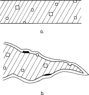
Fig. 1
(a) An ideal specimen which is flat, parallel-sided and undistorted, (b) Ạ more usual specimen which tapers, droops and is covered with a layer of oxide, contaminant, and dirt. Some of its second-phase particles have been etched out and some stand proud of the foil.
2
Initial preparation of sheet or disc
The thinning of bulk mater...
Table of contents
- Cover
- Title Page
- Copyright Page
- Contents
- 1 The specimen: what we are trying to achieve
- 2 Initial preparation of sheet or disc
- 3 Final thinning
- 4 Replicas
- 5 Mounting and storing specimens
- Appendix. Materials and suppliers
- References
- Index
Frequently asked questions
Yes, you can cancel anytime from the Subscription tab in your account settings on the Perlego website. Your subscription will stay active until the end of your current billing period. Learn how to cancel your subscription
No, books cannot be downloaded as external files, such as PDFs, for use outside of Perlego. However, you can download books within the Perlego app for offline reading on mobile or tablet. Learn how to download books offline
We are an online textbook subscription service, where you can get access to an entire online library for less than the price of a single book per month. With over 1.5 million books across 990+ topics, we’ve got you covered! Learn about our mission
Look out for the read-aloud symbol on your next book to see if you can listen to it. The read-aloud tool reads text aloud for you, highlighting the text as it is being read. You can pause it, speed it up and slow it down. Learn more about Read Aloud
Yes! You can use the Perlego app on both iOS and Android devices to read anytime, anywhere — even offline. Perfect for commutes or when you’re on the go.
Please note we cannot support devices running on iOS 13 and Android 7 or earlier. Learn more about using the app
Please note we cannot support devices running on iOS 13 and Android 7 or earlier. Learn more about using the app
Yes, you can access Specimen Preparation for Transmission Electron Microscopy of Materials by PJ Goodhew in PDF and/or ePUB format, as well as other popular books in Technology & Engineering & Biology. We have over 1.5 million books available in our catalogue for you to explore.