This practical resource shows the clinician how to handle and use the tools, devices, and implants required for endovascular interventions. A firm foundation in understanding the purpose, technical parameters, and function of each tool is required by vascular surgeons, interventional cardiologists, interventional radiologists, and other vascular specialists. Medical students, trainees, and physicians starting in practice will enhance their endovascular knowledge, which will allow for improved and seamless patient care. Even the experienced practitioner may find new tips in this book!
eBook - ePub
Endovascular Tools and Techniques Made Easy
- 132 pages
- English
- ePUB (mobile friendly)
- Available on iOS & Android
eBook - ePub
Endovascular Tools and Techniques Made Easy
About this book
Trusted by 375,005 students
Access to over 1 million titles for a fair monthly price.
Study more efficiently using our study tools.
Information
Topic
MedicineSubtopic
CardiologyCHAPTER 1
INTRODUCTION TO THE ENDOVASCULAR SUITE AND BASIC PRINCIPLES OF ANGIOGRAPHY
Jason Ty Turner, Virginia L. Wong
- Initial Procedural Workflow
- Procedural Imaging Step-by-Step
- Post-Procedure Workflow
- References
Learning Objectives
- Become familiar with typical equipment, personnel, devices, and workflow utilized in a Cardiac Catheterization Laboratory or a hybrid operating room
- Become familiar with nomenclature and basic concepts of fluoroscopy
- Understand the basic principles of angiography and radiation safety
Recently, there have been rapid advancements in endovascular technologies to treat arterial, venous, neurologic, and cardiac pathologies. These interventions commonly take place in an endovascular suite, such as a Cardiac Catheterization Lab (commonly referred to as the “Cath Lab”) or a hybrid operating room equipped with fixed (permanently mounted) fluoroscopic imaging equipment. Other venues with similar equipment are “Interventional Radiology Special Procedures” area, and outpatient angiography areas (office-based lab/OBL, ambulatory surgery center/ASC, etc.). Interventions can be performed at body sites remote from the point of vascular access using fluoroscopy to guide the real-time passage of wires, catheters, and devices to those sites for treatment. For simplicity, we will use endovascular suite as a generic term to encompass all venues where endovascular procedures, including angiography and intervention, can be performed.
A number of team members work together in the endovascular suite, and each has a specific role and responsibilities:
- Interventionalist (or Operator): A physician specialist trained to perform endovascular procedures and responsible for the conduct of the case, safety processes, supervision of other team members, and management of complications. Often, the interventionalist directs patient sedation, while a separate individual, such as a circulating nurse or an anesthetist, monitors patient comfort and vital signs during the case.
- Conscious Sedation Nurse: A nurse specialist who may manage all aspects of patient sedation and monitoring. In some venues (such as hybrid operating room [OR]) an anesthesiologist may provide sedation, particularly for more complex cases.
- Circulating Nurse: This individual monitors the patient's condition, gathers supplies and opens equipment onto the sterile field, and performs point-of-care testing (e.g., blood glucose, activated clotting time) when needed.
- Radiation Technologist: A trained and licensed individual who operates fluoroscopy equipment and assists the interventionalist in handling wires, catheters, and devices during the case.
- Recording Nurse or Tech: This individual monitors the entire procedure from a separate control room, documenting the timing and conduct of each procedure step, the equipment used, and other important procedural parameters. A recording nurse or tech more often assists an interventionalist in a Cath Lab setting.
Generally, all team members assist with loading the patient onto the table and preparing to start the case, as well as with patient transfer to the recovery room afterward, and then room turnover for the following case. Good teamwork and clear communication are essential for maintaining a smooth workflow and safe environment for the patients and staff during the procedure.
Initial Procedural Workflow
Patients are typically prepared for their procedure in a pre-procedural area. Medical history, vital signs, and pertinent lab values are reviewed; consent for the procedure is obtained; and intravenous access secured. The patient is then brought to the endovascular suite (Figure 1.1) and placed on the procedure table. Hemodynamic and cardiorespiratory monitoring are established and supplemental oxygen is provided. Following institutional pre-procedural patient identification protocols, the appropriate body part is exposed, prepped into a sterile field, and intravenous sedation is administered.
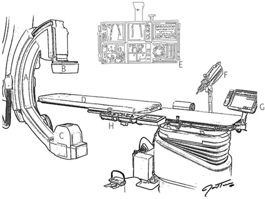
Figure 1.1Key components of the endovascular suite. (A) C-arm. (B) Image intensifier (I-I). (C) X-ray source. (D) Procedure table. (E) Display screen. (F) Power injector. (G) Control panel for imaging system. (H) Tableside control for maneuvering table and C-arm.
Vascular access is most often achieved percutaneously with the modified Seldinger technique, using ultrasound or fluoroscopy to guide introduction of a needle into the vessel lumen. Surgical exposure of the access vessel via cut down approach can also be employed and is common during combined open and endovascular procedures performed in a hybrid OR. A guidewire is advanced through the access needle that serves as a rail, or pathway, for other devices such as sheaths and catheters to follow. These items can be advanced along the wire to various distant sites for intervention, using fluoroscopy to visualize their progress and position inside the body.
Procedural Imaging Step-by-Step
- Typically, fluoroscopy is utilized for visualizing through body tissues and structures. Stepping on a foot pedal activates the imaging system. A single fluoroscopic image captured by simple press-and-release of the pedal is referred to as a spot image.
- Blood vessels and the blood contained within them have the same radiodensity as other soft tissues, and so are not visualized with plain fluoroscopy. Therefore, radiodense contrast dye is administered into the vessel lumen to provide differentiation from surrounding tissues and allow them to be seen. There are a number of common contrast agents with varying characteristics, rendering each particularly useful for certain applications (see Chapter 4, “Contrast Agents and Their Delivery”).
- Contrast dye may be either hand injected, using a syringe attached to an intraluminal catheter, or power injected, using a specific mechanical injector. A power injector is capable of administering much greater volumes of contrast at a higher rate and pressure than is possible by hand, and so is useful when trying to fill a large vessel or one with rapid blood flow (such as the aorta) with contrast. The power injector must be programmed with the desired rate of contrast administration (milliliters per second) and the total amount of contrast volume (mL) to be injected. This conventional programming sequence results in the common expression for communicating power injector settings to other team members, where “10 for 20” means that a 20 mL volume of contrast will be injected at a rate of 10 mL per second. Other injection parameters such as pressure, rise, and delay can be set separately.
- Prolonged pedal activation will capture and display multiple images using pulse dose fluoroscopy, with a frame (image capture) rate that is adjustable. A slower frame rate, such as 2–3 frames/second, is selected when using fluoroscopy to aid navigation of wires, catheters, and other devices. Imaging rapid movements or changes in position requires a higher frame rate. Cineangiography, or a cine run, is a series of successive X-ray images typically obtained at 15–30 frames/second and is used to capture the movement of radiopaque contrast injected into vessels.
- Digital subtraction angiography (DSA) uses an algorithm to digitally subtract out radiopaque structures from images obtained during a cine run. This is useful for obtaining isolated views of contrast-enhanced vessels without visual interference by adjacent or overlying bony structures and other radiodense materials such as orthopedic hardware, arterial calcification, metallic vascular stents, etc. The pre-contrast image, or mask, is obtained during the first few seconds of DSA exposure, then subtracted from subsequent frames of the same cine run. Contrast is then injected and appears radiodense on a background devoid of competing radiopaque structures. Road mapping is a technique used to overlay a DSA image of contrast-enhanced vessels on top of live pulse dose fluoroscopy, thus providing a live map for guidance of wires and devices in the vessel. Some advanced systems have the capability to pair live fluoroscopic imaging with pre-procedural computed tomography (CT) scans, further enhancing aided navigation in more complex procedures. The use of special fluoroscopy modes improves identification of vascular pathology and can help minimize the amount of radiation and contrast used during a procedure.
The fluoroscopy procedure table allows for movement of the patient in several axes (Figure 1.2). A mushroom-shaped knob on the tableside control panel is depressed to unlock and then “float” the tabletop over its support in the X and Y planes. A separate control raises or lowers the height of the tabletop in the Z plane, while tilted posi...
Table of contents
- Cover
- Half Title
- Title
- Copyright
- Dedication
- Contents
- Contributors
- Preface
- Chapter 1 Introduction to the Endovascular Suite and Basic Principles of Angiography
- Chapter 2 Pre-Procedural Risk Assessment and Optimization
- Chapter 3 Radiation Safety for You and Your Patient
- Chapter 4 Contrast Agents and Their Delivery
- Chapter 5 Vascular Access and Sheaths
- Chapter 6 Guidewires
- Chapter 7 Catheters
- Chapter 8 CTO Techniques and Re-Entry Devices
- Chapter 9 Angioplasty Balloons and Technique
- Chapter 10 Stents
- Chapter 11 Stent Grafts
- Chapter 12 Atherectomy Devices
- Chapter 13 Intravascular Imaging Modalities (OCT and IVUS)
- Chapter 14 Post-Procedural Hemostasis and Closure Devices
- Index
Frequently asked questions
Yes, you can cancel anytime from the Subscription tab in your account settings on the Perlego website. Your subscription will stay active until the end of your current billing period. Learn how to cancel your subscription
No, books cannot be downloaded as external files, such as PDFs, for use outside of Perlego. However, you can download books within the Perlego app for offline reading on mobile or tablet. Learn how to download books offline
Perlego offers two plans: Essential and Complete
- Essential is ideal for learners and professionals who enjoy exploring a wide range of subjects. Access the Essential Library with 800,000+ trusted titles and best-sellers across business, personal growth, and the humanities. Includes unlimited reading time and Standard Read Aloud voice.
- Complete: Perfect for advanced learners and researchers needing full, unrestricted access. Unlock 1.4M+ books across hundreds of subjects, including academic and specialized titles. The Complete Plan also includes advanced features like Premium Read Aloud and Research Assistant.
We are an online textbook subscription service, where you can get access to an entire online library for less than the price of a single book per month. With over 1 million books across 990+ topics, we’ve got you covered! Learn about our mission
Look out for the read-aloud symbol on your next book to see if you can listen to it. The read-aloud tool reads text aloud for you, highlighting the text as it is being read. You can pause it, speed it up and slow it down. Learn more about Read Aloud
Yes! You can use the Perlego app on both iOS and Android devices to read anytime, anywhere — even offline. Perfect for commutes or when you’re on the go.
Please note we cannot support devices running on iOS 13 and Android 7 or earlier. Learn more about using the app
Please note we cannot support devices running on iOS 13 and Android 7 or earlier. Learn more about using the app
Yes, you can access Endovascular Tools and Techniques Made Easy by Vikram S. Kashyap, Matthew Janko, Justin A. Smith, Vikram S. Kashyap,Matthew Janko,Justin A. Smith in PDF and/or ePUB format, as well as other popular books in Medicine & Cardiology. We have over one million books available in our catalogue for you to explore.