Digital images have several benefits, such as faster and inexpensive processing cost, easy storage and communication, immediate quality assessment, multiple copying while preserving quality, swift and economical reproduction, and adaptable manipulation. Digital medical images play a vital role in everyday life. Medical imaging is the process of producing visible images of inner structures of the body for scientific and medical study and treatment as well as a view of the function of interior tissues. This process pursues disorder identification and management.
Medical imaging in 2D and 3D includes many techniques and operations such as image gaining, storage, presentation, and communication. The 2D and 3D images can be processed in multiple dimensions. Depending on the requirement of a specific problem, one must identify various features of 2D or 3D images while applying suitable algorithms. These image processing techniques began in the 1960s and were used in such fields as space, clinical purposes, the arts, and television image improvement. In the 1970s, with the development of computer systems, the cost of image processing was reduced and processes became faster. In the 2000s, image processing became quicker, inexpensive, and simpler. In the 2020s, image processing has become a more accurate, more efficient, and self-learning technology.
This book highlights the framework of the robust and novel methods for medical image processing techniques in 2D and 3D. The chapters explore existing and emerging image challenges and opportunities in the medical field using various medical image processing techniques. The book discusses real-time applications for artificial intelligence and machine learning in medical image processing. The authors also discuss implementation strategies and future research directions for the design and application requirements of these systems.
This book will benefit researchers in the medical image processing field as well as those looking to promote the mutual understanding of researchers within different disciplines that incorporate AI and machine learning.
FEATURES
Highlights the framework of robust and novel methods for medical image processing techniques
Discusses implementation strategies and future research directions for the design and application requirements of medical imaging
Examines real-time application needs
Explores existing and emerging image challenges and opportunities in the medical field
Trusted by 375,005 students
Access to over 1.5 million titles for a fair monthly price.
1.2 Comparison between 2D and 3D Techniques in Medical Imaging
1.3 Importance of 3D Medical Image
1.4 Medical Imaging Types and Modalities
1.5 Computer Vision System Works in 3D Image Analysis
1.6 Various Techniques in 3D Image Processing in Medical Imaging
1.7 Types of Medical Imaging Compressed by 3D Medical Visualization
1.8 3D Ultrasound Shortens the Imaging Development
1.9 Conclusion
References
1.1Introduction
3D Image Analysis is the visualization, processing and evaluation of 3D photo statistics through geometric transformation, filtering, picture segmentation and other morphological operations. 3D conception forms the basis of contemporary radiology. 3D experimental imaging is a modern visual imaging scientific expertise that affords an enriched image of the interior body for scientific assessment making use of 3D imaging modalities. 3D scientific imaging provides more effective pictures of blood vessels and better images of bones. It is undisputable that 3 Dimensional (3D) imaging is continuously improving with the continued enhancement of instrumentation.
1.2Comparison Between 2D and 3D Techniques in Medical Imaging
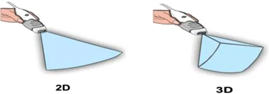
2D and 3D refer to the genuine dimensions in a computer workspace. 2D is “flat”; using horizontal and vertical (X and Y) dimensions; the image graph has solely two dimensions and turns into a line. 3D provides the depth (Z) dimension. This 0.33-dimension permits rotation and visualization from a couple of perspectives. It is in effect the distinction between an image and a sculpture.
For example, taking the pattern image graphs of echocardiography, there is the volumetric method to statistics acquisition in 2D and 3D (Figure 1.1).
Figure 1.1 2D (left panel) and 3D (right panel) Echocardiography Visualization Image.
Medical imaging has developed extensively since the early days of CT scanners and mammography equipment. With 3D scientific imaging, healthcare professionals were able to obtain access to fresh angles, resolutions, and small detail that provided an outstanding portrait of the physical section in query, at the same time as reducing the amount of radioactivity in patients [1, 2, 3]. In recent decades, the quantity of 3D scientific imaging has doubled in number every month to about one hundred thirty instances per day by 2018. The science of scanning has become a superior technology in creating statistical units that can make 3D images clearer with greater decision precision and much less noise and artifacts. Medical imaging has superior technological know-how in particular when it comes to these slice counts; it permits us to enlarge the precision of the pictures that we are shooting and, additionally signify the 3d mannequin of the anatomy, which used to be a substitute no longer feasible in the early days of the process (Figure 1.2).
Figure 1.2 Medical Imaging.
1.3Importance of 3D Medical Image
As we are all aware, medical imaging encompasses distinctive imaging modalities (a kind of technology used to gather structural or purposeful pictures of the body) such as radiography, ultrasound, nuclear prescription, computed tomography (CT), magnetic resonance and seen light. This requires techniques to image graph the body for diagnostictic and therapeutic purpose and performs an essential function in enhancing medical treatment. This proves that clinical imaging is regularly justified in the follow up of an ailment already recognized or treated [4, 5].
Medical imaging, in particular X-ray, primarily built investigations plus ultrasonography, stays necessary for a range of scientific putting and by altogether predominant stages of fitness precaution. In communal fitness and protective remedy by way of suitable as in each healing and relaxing care, good choices rely on the right analyses. However, medicinal/scientific decisions might also remain adequate former to therapy of numerous circumstances, the practise of diagnostic imaging offerings is dominant in confirming, efficiently measuring and authenticating publications of several ailments as nicely as in an evaluating reaction to treatment. Through accelerated health care coverage plus the growing accessibility of clinical apparatus, the range of world imaging-based strategies continues to grow significantly. Accurate and safe forms of imaging remain necessary in clinical practice and can reduce the use of pointless procedures. For instance, some medical interventions can be prevented if easy diagnostic imaging such as ultrasound is available.
It is well known that 3D picture processing is a tremendous tool for extent calculation, measurement, and quantitative analysis. It starts off evolved from 3D fashions of the patient, routinely recognized and extracted from anatomical structures, analysis, and surgical simulations can be supported. Moreover, with the usage of augmented actuality capabilities, it is feasible to merge preoperative or intraoperative information with reality, which is a precious device in the discipline of image guided surgery. 3D technological know-how has changed scientific imaging developing the opportunity for talent mapping with excessive decision microscopy. It has the capacity to discover character neurons, hint connections between them, and visualize organelles’ internal neurons.
The fundamental and first step in 3D image processing is the division of a picture which organizes pixels into substances or collections. 3D image division makes it practical to make 3D versions for more than one object and function with quantitative evaluation aimed at the extent, mass, and different factors of identified substances.
New images are taken, whether by CT, MRI, or microscopy image diagram as a 3D range of voxels/pixels. Individual voxel takes a greyscale vary from 0 to 65535 in the sixteen-bit pixel instance or 0 to 255 in the eight-bit pixel case. A segmented image, on the different hand, provides a less complicated explanation of substances that allows an introduction of 3D level methods or shows point data. When the fresh image graph is conveniently displayed as 3D evaluation, then imagining requires clearly described objective limits after growing models. Taking as an instance, to generate a 3D version of humanoid intelligence from an MRI image, the intelligence wishes to be recognized first inside the image graph and before its periphery manifest and used for 3D translation. The pixel recognition method remains known as image division, which recognises the qualities of pixels and describes the limitations for pixels that go to an identical group. Moreover, dimensions and numerical evaluation for restrictions such as region, boundary, quantity, and extent can be acquired effortlessly once objective limits are distinct.
1.4Medical Imaging Types and Modalities
Different kinds of medicinal imaging contain:
i.CT (Computed Tomography)
CT or CAT (pc axial tomography) images are the shape of X-ray that generates 3D images for analysis. It makes use of X-rays towards frame supply section images. A scanner with CT takes an outsized round establishing aimed at the affected person lying on a motorized desk. The X-ray delivers in addition a sensor then it rotates around the affected idividual, generating a slight ‘fan-shaped’ ray of X-rays that permits via part of the patient’s physique to make a picture. Those pictures are then assembled into single, or multiple images of interior organs and tissues. The CT scans supply higher transparency in comparison to traditional X-rays through greater unique picture graphs of the internal organs, bones, mild tissues, and blood vessels within the frame. The advantages of the use of CT scans outweigh the risks, which similar to X-rays, include cancer, damage to an unborn child, or allergic reaction to the chemicals in contrast material. In many instances, using a CT scan removes the need for experimental surgical operations. It is essential that when scanning children, the radiation dosage is lower than that used for adults. In many hospitals, a paediatrics CT scanner is available for that purpose.
ii.MRI (Magnetic Resonance Imaging)
MRI scans generate diagnostic image graphs without emission of dangerous radiation. Magnetic resonance Imaging (MRI) makes use of strong magnetic placed besides radio waves to produce pictures of the body which cannot be detected by X-rays or CT scans, i.e., it enables joints, ligaments and soft tissue to be visible [6, 7, 8]. The MRI is frequently used to observe interior detail to detect strokes, tumours, spinal cord accidents, aneurysms, and intelligence function. We realize the majority of the human body consists of water, and every water molecule consists of a hydrogen nucleus (proton) which become allied in a magnetic field. An MRI scanner provides a secure magnetic field to support the proton ‘spins’. A radio frequency is then applied which propels the protons to ‘flip’ their spins in advance than return to their proper arrangement. Protons in particular body organs revert to their regular spins at dissimilar rates so the MRI can differentiate among numerous types of tissue and detect any deformities. In what way the molecules ‘flip’ then arrive back at their ordinary spin arrangements is noted and processed into a picture. MRI doesn’t use ionizing radiation and is gradually being cast-off at some stage in pregnancy and not using a thing results at the unborn infant mentioned. But there are dangers related to using MRI scanning, and it isn’t endorsed as a primary analysis.Due to the strong magnets used, it is not suitable for individuals with any kind of steel implant, synthetic joints, and so on because of the chance they might be dislodged or heated up in the magnetic field.
iii.ULTRASOUND
Ultrasound remains the most secure method of scientific imaging and takes a large variety of packages. There aren’t any risk to the use of ultrasound, and it remains one of the best low-cost types of medical imaging available to us. Ultrasound makes use of sound waves instead of ionizing emission. High-frequency sound waves travel through the body with the aid of a transducer. Those waves then bounce back once they hit denser surfaces in the body and that is used to generate an image for prognosis. Another type of ultrasound often used is the ‘Doppler’ – an extraordinary method of the use of sound waves that allows the bloodflow via arteries and veins to be visible. Due to the absence of risk in ultrasound, it is the first choice of imaging in pregnancy. However, because its uses are considerable – emergency prognosis, cardiac, spine, and internal organs – it often is the first imaging option for patients.
iv.X-ray
X-ray imaging – X-ray consitutes the oldest and most used form of imaging; indeed, most people have had at least one X-ray in their life. In 1895, it was discovered that X-rays are a form of electromagnetic radiation. X-rays work on a wavelength and frequency that we are not able to view with the bare human eye, but it can...
Table of contents
Cover
Half Title
Title Page
Copyright Page
Contents
Preface
Introduction
Editors
Contributors
Chapter 1 An Introduction to Medical Image Analysis in 3D
Chapter 2 Automated Epilepsy Seizure Detection from EEG Signals Using Deep CNN Model
Chapter 3 Medical Image De-Noising Using Combined Bayes Shrink and Total Variation Techniques
Chapter 4 Detection of Nodule and Lung Segmentation Using Local Gabor XOR Pattern in CT Images
Chapter 5 Medical Image Fusion Using Adaptive Neuro Fuzzy Inference System
Chapter 6 Medical Imaging in Healthcare Applications
Chapter 7 Classification of Diabetic Retinopathy by Applying an Ensemble of Architectures
Chapter 8 Compression of Clinical Images Using Different Wavelet Function
Chapter 9 PSO-Based Optimized Machine Learning Algorithms for the Prediction of Alzheimer’s Disease
Chapter 10 Parkinson’s Disease Detection Using Voice Measurements
Chapter 11 Speech Impairment Using Hybrid Model of Machine Learning
Chapter 12 Advanced Ensemble Machine Learning Model for Balanced BioAssays
Chapter 13 Lung Segmentation and Nodule Detection in 3D Medical Images Using Convolution Neural Network
Index
Frequently asked questions
Yes, you can cancel anytime from the Subscription tab in your account settings on the Perlego website. Your subscription will stay active until the end of your current billing period. Learn how to cancel your subscription
No, books cannot be downloaded as external files, such as PDFs, for use outside of Perlego. However, you can download books within the Perlego app for offline reading on mobile or tablet. Learn how to download books offline
We are an online textbook subscription service, where you can get access to an entire online library for less than the price of a single book per month. With over 1.5 million books across 990+ topics, we’ve got you covered! Learn about our mission
Look out for the read-aloud symbol on your next book to see if you can listen to it. The read-aloud tool reads text aloud for you, highlighting the text as it is being read. You can pause it, speed it up and slow it down. Learn more about Read Aloud
Yes! You can use the Perlego app on both iOS and Android devices to read anytime, anywhere — even offline. Perfect for commutes or when you’re on the go. Please note we cannot support devices running on iOS 13 and Android 7 or earlier. Learn more about using the app
Yes, you can access Artificial Intelligence and Machine Learning in 2D/3D Medical Image Processing by Rohit Raja, Sandeep Kumar, Shilpa Rani, K. Ramya Laxmi, Rohit Raja,Sandeep Kumar,Shilpa Rani,K. Ramya Laxmi in PDF and/or ePUB format, as well as other popular books in Medicine & Computer Vision & Pattern Recognition. We have over 1.5 million books available in our catalogue for you to explore.