eBook - ePub
Imaging in Clinical Neurosciences for Non-radiologists
An Atlas
- 191 pages
- English
- ePUB (mobile friendly)
- Available on iOS & Android
eBook - ePub
Imaging in Clinical Neurosciences for Non-radiologists
An Atlas
About this book
This Atlas presents both normal and pathological conditions of the Brain and Spine pictorially. Targeted towards non-radiologists, it is a unique book with well labeled and self-explanatory images. All routine conditions involving neuroradiology have been included. Images from different radiological modalities such as X-ray, Computed Tomography (CT), Magnetic Resonance Imaging (MRI) and Digital Subtraction Angiography (DSA) have also been included. This book aims to serve as a ready reckoner for clinicians, trainees, residents as well as professional radiologists.
Key Features
- Discusses topics related to allied branches of neurology, neuroanesthesia, neurointensive care and neurosurgery
-
- Presents both common and uncommon neurological conditions
-
- Contains actual real-life scans and images
-
- Works as a unique, quick reference guide of neuroradiological images for non-radiologists
Frequently asked questions
Yes, you can cancel anytime from the Subscription tab in your account settings on the Perlego website. Your subscription will stay active until the end of your current billing period. Learn how to cancel your subscription.
At the moment all of our mobile-responsive ePub books are available to download via the app. Most of our PDFs are also available to download and we're working on making the final remaining ones downloadable now. Learn more here.
Perlego offers two plans: Essential and Complete
- Essential is ideal for learners and professionals who enjoy exploring a wide range of subjects. Access the Essential Library with 800,000+ trusted titles and best-sellers across business, personal growth, and the humanities. Includes unlimited reading time and Standard Read Aloud voice.
- Complete: Perfect for advanced learners and researchers needing full, unrestricted access. Unlock 1.4M+ books across hundreds of subjects, including academic and specialized titles. The Complete Plan also includes advanced features like Premium Read Aloud and Research Assistant.
We are an online textbook subscription service, where you can get access to an entire online library for less than the price of a single book per month. With over 1 million books across 1000+ topics, we’ve got you covered! Learn more here.
Look out for the read-aloud symbol on your next book to see if you can listen to it. The read-aloud tool reads text aloud for you, highlighting the text as it is being read. You can pause it, speed it up and slow it down. Learn more here.
Yes! You can use the Perlego app on both iOS or Android devices to read anytime, anywhere — even offline. Perfect for commutes or when you’re on the go.
Please note we cannot support devices running on iOS 13 and Android 7 or earlier. Learn more about using the app.
Please note we cannot support devices running on iOS 13 and Android 7 or earlier. Learn more about using the app.
Yes, you can access Imaging in Clinical Neurosciences for Non-radiologists by Hemanshu Prabhakar,S. Leve Joseph Devarajan in PDF and/or ePUB format, as well as other popular books in Medicine & Anesthesiology & Pain Management. We have over one million books available in our catalogue for you to explore.
Information
1
Normal anatomy

Figure 1.1Brain axial section—Pons level: (A) Computed tomography scan of brain in axial view at the level of pons showing normal structures. (B) Magnetic resonance imaging of brain in axial view at the level of pons showing normal structures.

Figure 1.2 Brain axial section—Midbrain level: (A) Computed tomography scan of brain in axial view at the level of midbrain and suprasellar cistern showing normal structures. (B) Magnetic resonance imaging of brain in axial view at the same level showing normal structures.
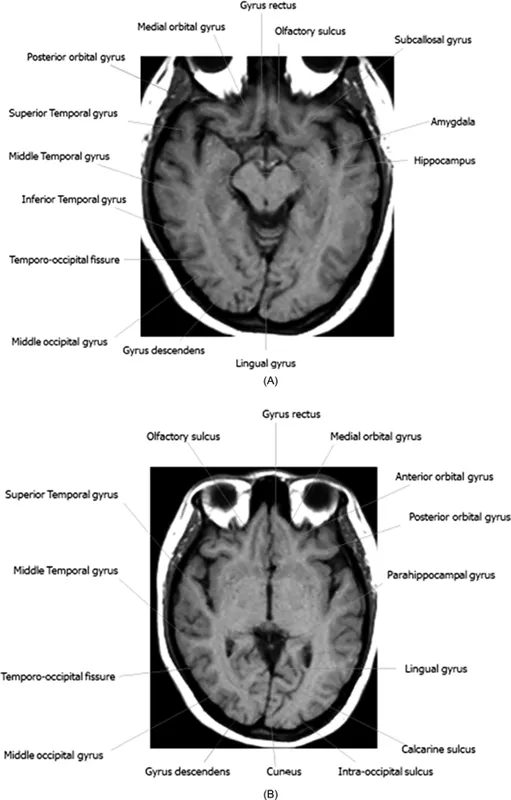
Figure 1.3 Brain axial section—Upper midbrain and lower III ventricle levels: (A and B) Magnetic resonance imaging of brain in axial view at the upper midbrain and lower third ventricle levels showing normal structures.
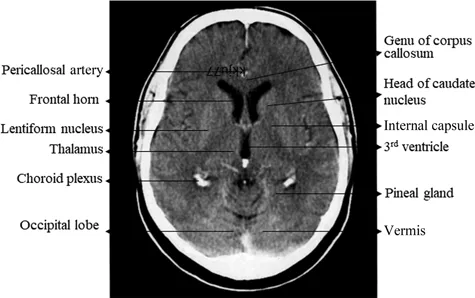
Figure 1.4 Brain axial section—Lower III ventricular level. Computed tomography scan of brain in axial view at the lower third ventricular level showing normal structures.
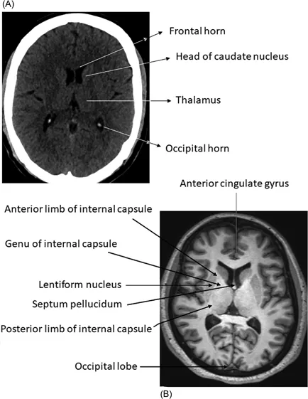
Figure 1.5 Brain axial section—Upper III ventricular level: (A) Computed tomography scan of brain in axial view at the upper third ventricular level showing normal structures. (B) Magnetic resonance imaging of brain in axial view at the upper third ventricular level showing normal structures.
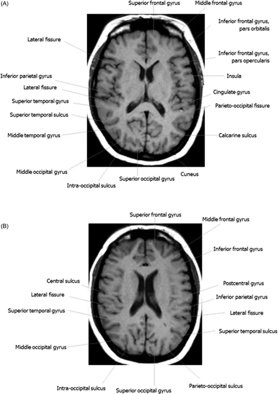
Figure 1.6 Brain axial section—Magnetic resonance imaging of brain in axial view at the upper third ventricular and body of the lateral ventricles levels respectively showing normal structures.
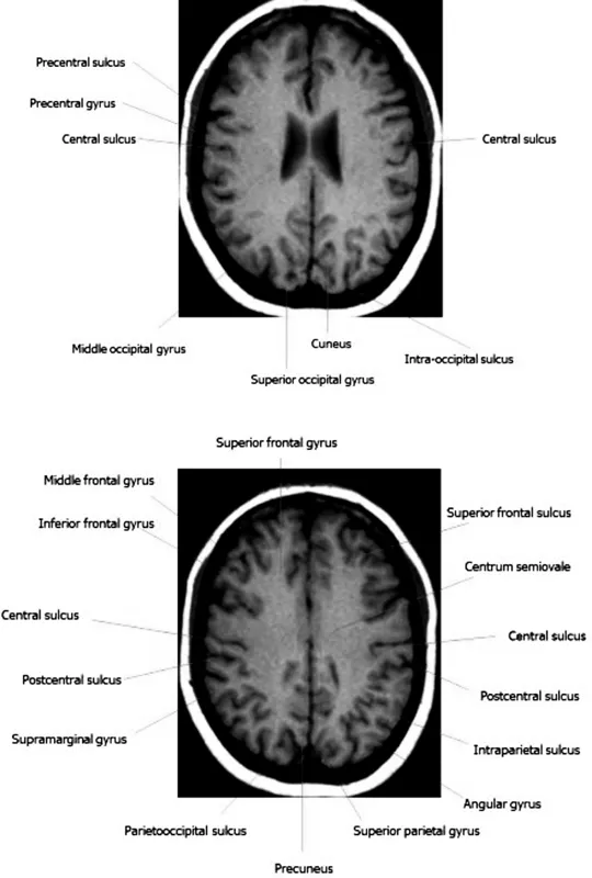
Figure 1.7 Brain axial section—Magnetic resonance imaging of brain in axial view at the centrum semiovale level showing normal structures.
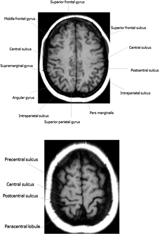
Figure 1.8 Brain axial section—High frontoparietal. Magnetic resonance imaging of brain in axial view at the high frontoparietal levels showing normal structures.
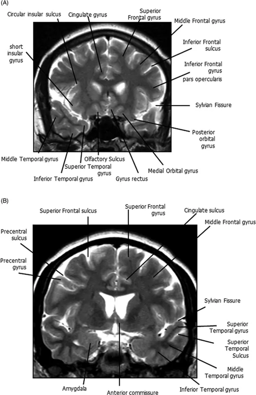
Figure 1.9 Brain coronal section—Frontal lobes level: (A) and (B) Magnetic resonance imaging of brain in coronal section at the level of frontal horns showing normal structures.
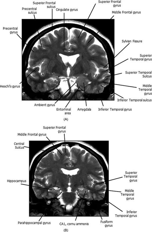
Figure 1.10 Brain coronal sections—Lateral ventricles level: (A) and (B) Magnetic resonance imaging of brain in coronal section at the level of body of lateral ventricles showing normal structures.
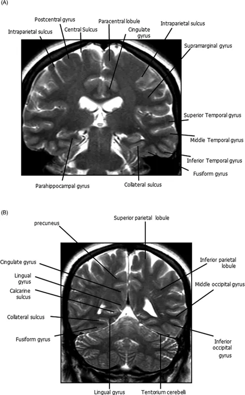
Figure 1.11 Brain coronal sections—Posterior lateral ventricles and occipital horns level: (A) Magnetic resonance imaging of brain in coronal section at the level of posterior lateral ventricles showing normal structures. (B) Magnetic resonance imaging of brain in coronal section at the level of occipital horns showing normal structures.
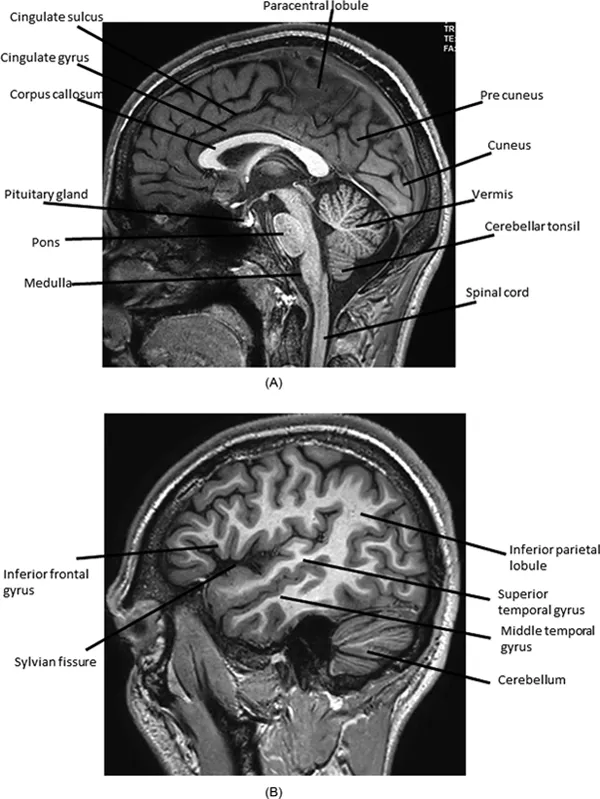
Figure 1.12 Brain sagittal section: (A) and (B) Magnetic resonance imaging of brain in mid-sagittal and parasagittal sections showing normal structures.
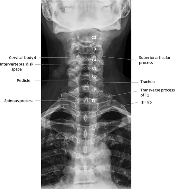
Figure 1.13 Spine X-ray of cervical spine—Anteroposterior view: X-ray of cervical spine in anteroposterior view showing normal vertebral structures.
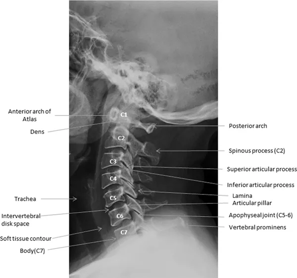
Figure 1.14 Spine X...
Table of contents
- Cover
- Half Title
- Title Page
- Copyright Page
- Dedication
- Contents
- Preface
- Acknowledgments
- Authors
- List of Abbreviations
- List of Contributors
- 1 Normal anatomy
- 2 Congenital anomalies
- 3 Epilepsy imaging
- 4 Neurocutaneous syndromes
- 5 Metabolic disorders
- 6 Demyelination and inflammatory disorders
- 7 Neurovascular diseases
- 8 Central nervous system tumors
- 9 Central nervous system infections
- 10 Neurotrauma
- 11 Degenerative brain and spine diseases
- Index