Handbook of Pediatric Hematology and Oncology
eBook - ePub

Handbook of Pediatric Hematology and Oncology

Children's Hospital and Research Center Oakland

Caroline A. Hastings, Joseph C. Torkildson, Anurag K. Agrawal

Share book
  1. English
  2. ePUB (mobile friendly)
  3. Available on iOS & Android
eBook - ePub

Handbook of Pediatric Hematology and Oncology

Children's Hospital and Research Center Oakland

Caroline A. Hastings, Joseph C. Torkildson, Anurag K. Agrawal

Book details
Book preview
Table of contents
Citations

About This Book

The revised guide to the diagnosis, management, and treatment of blood disorders and cancer in children

Children with blood disorders and cancer are a unique population that require specialized diagnostic considerations and management. The Handbook of Pediatric Hematology and Oncology has been designed to provide clinicians of all levels with practical guidance through an up-to-date algorithmic approach to these conditions. Assembled by a team of experts from the world-class Children's Hospital & Research Center Oakland, this updated third edition:

  • Presents up-to-date management and treatment guidelines for the most common pediatric blood disorders and malignancies
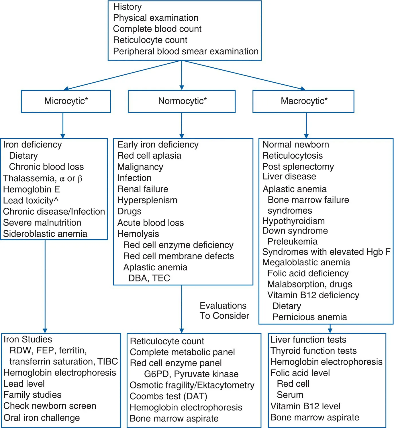
  • Provides an updated algorithmic approach for the diagnosis and management of the most common conditions and suggested readings
  • Utilizes rapid-referral tables containing visual representations of symptoms, lab findings, differentials, and treatment guidance
  • Incorporates case studies covering different hematologic and oncologic conditions, such as hemolytic anemia, sickle cell disease, hemophilia, neuroblastoma, and sarcomas of the soft tissue and bone
  • Includes a useful formulary that lists chemotherapy agents, dosing, mechanism of action, pregnancy category, indications, and side effects
  • Covers transfusion medicine, stem cell transplantation, management of central venous catheters, acute pain management, oncologic emergencies, and chemotherapy basics

With its direct guidance and portable design, the Handbook of Pediatric Hematology and Oncology: Children's Hospital & Research Center Oakland, Third Edition, will prove an invaluable resource to medical students, trainees and residents, pediatric hematology and oncology nurses, pediatricians, and early-career providers in pediatric hematology/oncology.

Frequently asked questions

How do I cancel my subscription?
Simply head over to the account section in settings and click on “Cancel Subscription” - it’s as simple as that. After you cancel, your membership will stay active for the remainder of the time you’ve paid for. Learn more here.
Can/how do I download books?
At the moment all of our mobile-responsive ePub books are available to download via the app. Most of our PDFs are also available to download and we're working on making the final remaining ones downloadable now. Learn more here.
What is the difference between the pricing plans?
Both plans give you full access to the library and all of Perlego’s features. The only differences are the price and subscription period: With the annual plan you’ll save around 30% compared to 12 months on the monthly plan.
What is Perlego?
We are an online textbook subscription service, where you can get access to an entire online library for less than the price of a single book per month. With over 1 million books across 1000+ topics, we’ve got you covered! Learn more here.
Do you support text-to-speech?
Look out for the read-aloud symbol on your next book to see if you can listen to it. The read-aloud tool reads text aloud for you, highlighting the text as it is being read. You can pause it, speed it up and slow it down. Learn more here.
Is Handbook of Pediatric Hematology and Oncology an online PDF/ePUB?
Yes, you can access Handbook of Pediatric Hematology and Oncology by Caroline A. Hastings, Joseph C. Torkildson, Anurag K. Agrawal in PDF and/or ePUB format, as well as other popular books in Medicine & Hematology. We have over one million books available in our catalogue for you to explore.

Information

Year
2021
ISBN
9781119210757
Edition
3
Subtopic
Hematology

1
Approach to the Anemic Child

Anemia is the condition in which the concentration of hemoglobin or the red cell mass is reduced below normal. Anemia results in a physiological decrease in the oxygen‐carrying capacity of the blood and reduced oxygen supply to the tissues. Causes of anemia are increased loss or destruction of red blood cells (RBCs) or a significant decreased rate of production. When evaluating a child with anemia, it is important to determine if the problem is isolated to one cell line (e.g., RBCs) or multiple cell lines (i.e., RBCs, white blood cells [WBCs], or platelets). When two or three cell lines are affected, it may indicate bone marrow involvement (e.g., leukemia, metastatic disease, and aplastic anemia), sequestration (i.e., hypersplenism), immune deficiency, or an immune‐mediated process (e.g., hemolytic anemia and immune thrombocytopenic purpura).

Evaluation of anemia

The evaluation of anemia includes a complete medical history, family history, physical examination, and laboratory assessment (see Figure 1.1).
The diagnosis of anemia is made after reference to established normal controls for age (Table 1.1). The blood smear and red cell indices are very helpful in the diagnosis and classification of anemia. They allow for classification by the cell size (mean corpuscular volume [MCV]), give the distribution of cell size (red cell distribution width [RDW]), and may give important diagnostic clues if specific morphological abnormalities are present (e.g., sickle cells, target cells, and spherocytes). The MCV, RDW, and reticulocyte count are helpful in the differential diagnosis of anemia. A high RDW, or anisocytosis, is seen in stress erythropoiesis and is often suggestive of iron deficiency or hemolysis. A normal or low reticulocyte count is an inappropriate response to anemia and suggests impaired red cell production. An elevated reticulocyte count suggests blood loss, hemolysis, or sequestration.
The investigation of anemia requires the following steps:
  1. The medical history of the anemic child (Table 1.2), as certain historical points may provide clues as to the etiology of the anemia.
  2. Detailed physical examination (Table 1.3), with particular attention to acute and chronic effects of anemia.
  3. Evaluation of the complete blood count (CBC), RBC indices, and peripheral blood smear, with classification by MCV, reticulocyte count, and RBC morphology. Consideration should also be given to the WBC and platelet counts as well as their respective morphologies.
    Schematic illustration of the diagnostic approach to the child with anemia.
    Figure 1.1 Diagnostic approach to the child with anemia (abbreviations: DBA, Diamond–Blackfan anemia; TEC, transient erythroblastopenia of childhood; RDW, red cell distribution width; FEP, free erythrocyte protoporphyrin; TIBC, total iron‐binding capacity; G6PD, glucose‐6‐phosphate dehydrogenase deficiency; DAT, direct antiglobulin test).*Refer to Table 1.1 for age‐based normal values.^Microcytosis with lead toxicity has been noted secondary to concomitant iron deficiency; see text.
  4. Determination of an etiology of the anemia by additional studies as needed (see Figures 1.11.3).

Interventions

Oral iron challenge

An oral iron challenge may be indicated in the patient with significant iron depletion, as documented by moderate‐to‐severe anemia and deficiencies in circulating and storage iron forms (such as an elevated total iron‐binding capacity [TIBC], low serum iron, low transferrin saturation, and low ferritin). Iron absorption is impaired in certain chronic disorders (autoimmune diseases such as systemic lupus erythematosus, peptic ulcer disease, ulcerative colitis, and Crohn's disease), by certain medications (antacids and histamine‐2 blockers), and by environmental factors such as lead toxicity.
Table 1.1 Red blood cell values at various ages.*
Age Hemoglobin (g/dL) MCV (fL)
Mean −2 SD Mean −2 SD
Birth (cord blood) 16.5 13.5 108 98
1–3 d (capillary) 18.5 14.5 108 95
1 wk 17.5 13.5 107 88
2 wk 16.5 12.5 105 86
1 mo 14.0 10.0 104 85
2 mo 11.5 9.0 96 77
3–6 mo 11.5 9.5 91 74
0.5–2 y 12.0 11.0 78 70
2–6 y 12.5 11.5 81 75
6–12 y 13.5 11.5 86 77
12–18 y female 14.0 12.0 90 78
12–18 y male 14.5 13.0 88 78
18–49 y female 14.0 12.0 90 80
18–49 y male 15.5 13.5 90 80
* Compiled from the following sources: Dutcher TF. Lab Med 2:32–35, 1971; Koerper MA, et al. J Pediatr 89:580–583, 1976; Marner T. Acta Paediatr Scand 58:363–368,...

Table of contents