- 411 pages
- English
- ePUB (mobile friendly)
- Available on iOS & Android
eBook - ePub
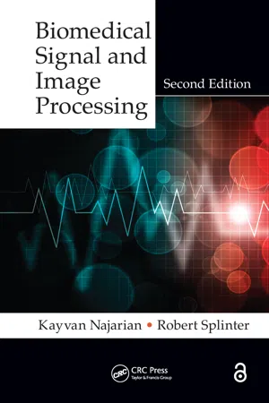
Biomedical Signal and Image Processing
About this book
Written for senior-level and first year graduate students in biomedical signal and image processing, this book describes fundamental signal and image processing techniques that are used to process biomedical information. The book also discusses application of these techniques in the processing of some of the main biomedical signals and images, such as EEG, ECG, MRI, and CT. New features of this edition include the technical updating of each chapter along with the addition of many more examples, the majority of which are MATLAB based.
Tools to learn more effectively

Saving Books

Keyword Search

Annotating Text

Listen to it instead
Information
Part I
Introduction to Digital Signal and Image Processing
1 Signals and Biomedical Signal Processing
1.1 INTRODUCTION AND OVERVIEW
The most fundamental concept that is frequently used in this book is a “signal.” It is imperative to clearly define this concept and to illustrate different types of signals encountered in signal and image processing. In this chapter, different types of signals are defined, and the fundamental concepts of signal transformation and processing are presented while avoiding detailed mathematical formulations.
1.2 WHAT IS A “SIGNAL”?
The definition of a signal plays an important role in understanding the capabilities of signal processing. We start this chapter with the definition of one-dimensional (1-D) signals. A 1-D signal is an ordered sequence of numbers that describes the trends and variations of a quantity. The consecutive measurements of a physical quantity taken at different times create a typical signal encountered in science and engineering. The order of the numbers in a signal is often determined by the order of measurements (or events) in “time.” A sequence of body temperature recordings collected in consecutive days forms an example of a 1-D signal in time. The characteristics of a signal lie in the order of the numbers as well as the amplitude of the recorded numbers, and the main task of all signal processing tools is to analyze the signal in order to extract important knowledge that may not be clearly visible to the human eyes.
We have to emphasize the point that not all 1-D signals are necessarily ordered in time. As an example, consider the signal formed by the recordings of the temperature simultaneously measured at different points along a metal rod where the distance from one end of the rod defines the order of the sequence. In such a signal, the points that are closer to the origin (one end of the metal rod) appear earlier in the sequence, and, as a result, the concept that orders the sequence is “distance in space” as opposed to time. However, due to abundance of time signals in many areas of science, in the literature of signal processing, the word “time” is often used to describe the axis that identifies order. In this book, without losing the generality of the results or concepts, we use the concept of time as the ordering axis, knowing that, in some signals, time should be replaced by other concepts such as space.
Many examples of biological 1-D signals are heavily used in medicine and biology. Recording of the electrical activities of the heart muscles, called electrocardiogram (ECG), is widely considered as the main diagnostic signal in assessment of the cardiovascular system. Electroencephalogram (EEG) is a signal that records the electrical activities of the brain and is heavily used in diagnostics of the central nervous system (CNS).
Multidimensional signals are simply extensions of the 1-D signals mentioned earlier, i.e., a multidimensional signal is a multidimensional sequence of numbers ordered in all dimensions. For example, an image is a two-dimensional (2-D) sequence of data where numbers are ordered in both dimensions. In almost all images, the numbers are ordered in space (for both dimensions). In a gray-scale image, the value of the signal for a given set of coordinates (x, y), i.e., g(x, y), identifies the image brightness level at those coordinates. There are several important types of image modalities that are heavily used for clinical diagnostics among which magnetic resonance imaging (MRI), computed tomography (CT), ultrasonic images, and positron emission tomography (PET) are the most commonly used ones. These imaging systems will be introduced in separate chapters dedicated to each image modality.
1.3 ANALOG, DISCRETE, AND DIGITAL SIGNALS
Based on the continuity of a signal in time and amplitude axes, the following three types of signals can be recognized:
1.3.1 ANALOG SIGNALS
These signals are co...
Table of contents
- Cover
- Half Title
- Title Page
- Copyright Page
- Dedication
- Table of Contents
- Preface
- Acknowledgments
- Introduction
- Part I Introduction to Digital Signal and Image Processing
- Part II Processing of Biomedical Signals
- Part III Processing of Biomedical Images
- Index
Frequently asked questions
Yes, you can cancel anytime from the Subscription tab in your account settings on the Perlego website. Your subscription will stay active until the end of your current billing period. Learn how to cancel your subscription
No, books cannot be downloaded as external files, such as PDFs, for use outside of Perlego. However, you can download books within the Perlego app for offline reading on mobile or tablet. Learn how to download books offline
Perlego offers two plans: Essential and Complete
- Essential is ideal for learners and professionals who enjoy exploring a wide range of subjects. Access the Essential Library with 800,000+ trusted titles and best-sellers across business, personal growth, and the humanities. Includes unlimited reading time and Standard Read Aloud voice.
- Complete: Perfect for advanced learners and researchers needing full, unrestricted access. Unlock 1.4M+ books across hundreds of subjects, including academic and specialized titles. The Complete Plan also includes advanced features like Premium Read Aloud and Research Assistant.
We are an online textbook subscription service, where you can get access to an entire online library for less than the price of a single book per month. With over 1 million books across 990+ topics, we’ve got you covered! Learn about our mission
Look out for the read-aloud symbol on your next book to see if you can listen to it. The read-aloud tool reads text aloud for you, highlighting the text as it is being read. You can pause it, speed it up and slow it down. Learn more about Read Aloud
Yes! You can use the Perlego app on both iOS and Android devices to read anytime, anywhere — even offline. Perfect for commutes or when you’re on the go.
Please note we cannot support devices running on iOS 13 and Android 7 or earlier. Learn more about using the app
Please note we cannot support devices running on iOS 13 and Android 7 or earlier. Learn more about using the app
Yes, you can access Biomedical Signal and Image Processing by Kayvan Najarian,Robert Splinter in PDF and/or ePUB format, as well as other popular books in Computer Science & Digital Media. We have over one million books available in our catalogue for you to explore.