
eBook - ePub
Making Sense of the ECG
A Hands-On Guide
Andrew Houghton
This is a test
Share book
- 244 pages
- English
- ePUB (mobile friendly)
- Available on iOS & Android
eBook - ePub
Making Sense of the ECG
A Hands-On Guide
Andrew Houghton
Book details
Book preview
Table of contents
Citations
About This Book
Interpreting an ECG correctly and working out what to do next can seem like a daunting task to the non-specialist, yet it is a skill that will be invaluable to any doctor, nurse or paramedic when evaluating the condition of a patient. Making Sense of the ECG has been written specifically with this in mind, and will help the student and more experienced healthcare practitioner to identify and answer crucial questions. This popular, easy-to-read and easy-to-remember guide to the ECG as a tool for diagnosis and management has been fully updated in its fifth edition to reflect the latest guidelines.
Frequently asked questions
How do I cancel my subscription?
Can/how do I download books?
At the moment all of our mobile-responsive ePub books are available to download via the app. Most of our PDFs are also available to download and we're working on making the final remaining ones downloadable now. Learn more here.
What is the difference between the pricing plans?
Both plans give you full access to the library and all of Perlego’s features. The only differences are the price and subscription period: With the annual plan you’ll save around 30% compared to 12 months on the monthly plan.
What is Perlego?
We are an online textbook subscription service, where you can get access to an entire online library for less than the price of a single book per month. With over 1 million books across 1000+ topics, we’ve got you covered! Learn more here.
Do you support text-to-speech?
Look out for the read-aloud symbol on your next book to see if you can listen to it. The read-aloud tool reads text aloud for you, highlighting the text as it is being read. You can pause it, speed it up and slow it down. Learn more here.
Is Making Sense of the ECG an online PDF/ePUB?
Yes, you can access Making Sense of the ECG by Andrew Houghton in PDF and/or ePUB format, as well as other popular books in Medicine & Medical Theory, Practice & Reference. We have over one million books available in our catalogue for you to explore.
Information
Chapter 1
Anatomy and physiology
The heart is a hollow muscular organ that pumps blood around the body. With each beat, it pumps, at rest, about 70 millilitres of blood and considerably more during exercise. Over a 70-year life span and at a rate of around 70 beats per minute, the heart will beat over 2.5 billion times.
The heart consists of four main chambers (left and right atria, and left and right ventricles) and four valves (aortic, mitral, pulmonary and tricuspid). Venous blood returns to the right atrium via the superior and inferior vena cavae, and leaves the right ventricle for the lungs via the pulmonary artery. Oxygenated blood from the lungs returns to the left atrium via the four pulmonary veins, and leaves the left ventricle via the aorta (Figure 1.1).
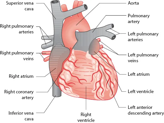
Figure 1.1 Cardiac anatomy.
Key point:
• The heart and major vessels.
The heart is made up of highly specialized cardiac muscle comprising myocardial cells (myocytes), which differs markedly from skeletal muscle because heart muscle:
•Is under the control of the autonomic nervous system
•Contracts in a repetitive and rhythmic manner
•Has a large number of mitochondria which make the myocytes resistant to fatigue
•Cannot function adequately in anaerobic (ischaemic) conditions
Cardiac Activation
Myocytes are essentially contractile but are capable of generating and transmitting electrical activity. Myocytes are interconnected by cytoplasmic bridges or syncytia, so once one myocyte cell membrane is activated (depolarized), a wave of depolarization spreads rapidly to adjacent cells.
Myocardial cells are capable of being:
•Pacemaker cells: These are found primarily in the sinoatrial (SA) node and produce a spontaneous electrical discharge.
•Conducting cells: These are found in:
•The atrioventricular (AV) node
•The bundle of His and bundle branches
•The Purkinje fibres
•Contractile cells: These form the main cell type in the atria and ventricles.
All myocytes are self-excitable with their own intrinsic contractile rhythm. Cardiac cells in the SA node located high up in the right atrium generate action potentials or impulses at a rate of about 60–100 per minute, a slightly faster rate than cells elsewhere such as the AV node (typically 40–60 per minute) or the ventricular conducting system (30–40 per minute), so the SA node becomes the heart pacemaker, dictating the rate and timing of action potentials that trigger cardiac contraction, overriding the potential of other cells to generate impulses. However, should the SA node fail or an impulse not reach the ventricles, cardiac contraction may be initiated by these secondary sites (‘escape rhythms’).
The Cardiac Action Potential
The process of triggering cardiac cells into function is called cardiac excitation–contraction coupling. Cells remain in a resting state until activated by changes in voltage due to the complex movement of sodium, potassium and calcium across the cell membrane (Figure 1.2); these are similar to changes which occur in nerve cells.
Phase 4: At rest, there is little spontaneous depolarization as the Na+/K+/ATPase pump maintains a negative stable resting membrane potential of about –90 mV. Some cardiac cells display automaticity or spontaneous regular action potentials, which generates action potentials in adjacent cells linked by cytoplasmic bridges or syncytia, so once one myocyte cell membrane is activated (depolarized), a wave of excitation spreads rapidly to adjacent cells; the SA node, whose cells are relatively permeable to sodium resulting in a less negative resting potential of about –55 mV, is usually the source of spontaneous action potentials.
Phase 0: There is rapid opening of sodium channels with movement of sodium into the cell, the resulting electrochemical gradient leading to a positive resting membrane potential.
Phase 1: When membrane potential is at its most positive, the electrochemical gradient causes potassium outflow and closure of sodium channels.
Phase 2: A plateau phase follows, with membrane ...