Radiation Detection: Concepts, Methods, and Devices provides a modern overview of radiation detection devices and radiation measurement methods. The book topics have been selected on the basis of the authors' many years of experience designing radiation detectors and teaching radiation detection and measurement in a classroom environment.
This book is designed to give the reader more than a glimpse at radiation detection devices and a few packaged equations. Rather it seeks to provide an understanding that allows the reader to choose the appropriate detection technology for a particular application, to design detectors, and to competently perform radiation measurements. The authors describe assumptions used to derive frequently encountered equations used in radiation detection and measurement, thereby providing insight when and when not to apply the many approaches used in different aspects of radiation detection. Detailed in many of the chapters are specific aspects of radiation detectors, including comprehensive reviews of the historical development and current state of each topic. Such a review necessarily entails citations to many of the important discoveries, providing a resource to find quickly additional and more detailed information.
This book generally has five main themes:
Physics and Electrostatics needed to Design Radiation Detectors
Properties and Design of Common Radiation Detectors
Description and Modeling of the Different Types of Radiation Detectors
Radiation Measurements and Subsequent Analysis
Introductory Electronics Used for Radiation Detectors
Topics covered include atomic and nuclear physics, radiation interactions, sources of radiation, and background radiation. Detector operation is addressed with chapters on radiation counting statistics, radiation source and detector effects, electrostatics for signal generation, solid-state and semiconductor physics, background radiations, and radiation counting and spectroscopy. Detectors for gamma-rays, charged-particles, and neutrons are detailed in chapters on gas-filled, scintillator, semiconductor, thermoluminescence and optically stimulated luminescence, photographic film, and a variety of other detection devices.
Trusted by 375,005 students
Access to over 1.5 million titles for a fair monthly price.
Great discoveries are made accidentally less often than the populace likes to think.
Wilhelm Röntgen
1.1A Brief History of Radiation Discovery
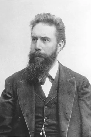
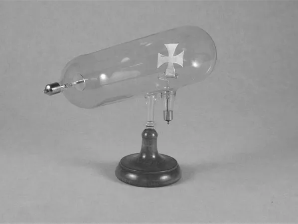
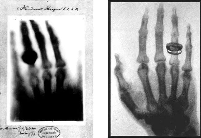
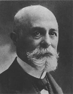
In 1895, within the town of Strasbourg (then located in Germany, but now in France), Wilhelm Conrad Röntgen (see Fig. 1.1) made the first observation of mysterious penetrating rays. For many years, these mysterious rays were called “Röntgen rays”; however, Röntgen actually named them“x rays,” as we know them today, after the common algebraic symbol for an unknown. In the famous experiment, Röntgen was operating a type of Crookes tube (see Fig. 1.2) while performing experiments with “cathode rays.” With the Crookes tube covered with black paper, he noticed that a plate coated with barium-platinocyanide (BaPt(CN)4), located approximately six feet away in the darkened room, was glowing. By placing objects with varying densities and thicknesses between the Crookes tube and the plate, he deduced that the mysterious emanations originated from the Crookes tube and that they were attenuated according to mass density and thickness of the absorber. A short while later, it was discovered that x rays are actually a form of electromagnetic radiation emitted from accelerated charged particles (electrons in Röntgen’s experiment). Röntgen also used photographic plates for his studies after he learned that film emulsions were exposed by the x rays. After experimenting with many x-ray image exposures of inanimate objects, perhaps the most famous x-ray photograph he developed is traditionally believed to be an image of his wife’s hand, Bertha Röntgen (see Fig. 1.3). Indeed photographic film is a type of radiation detector, and the x-ray image of Bertha Röntgen’s hand marks the beginning of medical imaging. For his discovery of x rays, Röntgen became the recipient in 1901 of the first Nobel Prize in Physics. These discoveries were made with two types of radiation detectors, namely the scintillating BaPt(CN)4-coated plate and the photographic plates.
Figure 1.1. Wilhelm Conrad Röntgen (1845–1923).
Figure 1.2. A Crookes tube was used as the x-ray source in Röntgen’s experiments. The Maltese cross in the vacuum tube is connected to a hinge that allows it to be flipped up or down.
Figure 1.3. (Left) Traditionally labeled as Bertha Röntgen’s hand, and probably the case, it actually was not labeled as such at the time of publication. (Right) Often mistakenly labeled as Bertha Röntgen’s hand, this image taken by Röntgen is actually that of Alfred von Kölliker’s hand. The discovery that x rays can be usedto produce images of bone structure marks the beginning of the modern medical imaging profession.
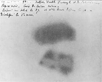
In 1896, shortly after the announcement of the discovery of x rays, Henri Becquerel (Fig. 1.4), while conducting experiments with his father’s geology samples, discovered natural radiation emissions. It was already known that many substances could be made to fluoresce by exposing them to visible light, and Becquerel was studying the fluorescence of sulphates of uranium and potassium by exposing his samples to sunlight and measuring the light emissions with photographic plates. In one instance, he prepared a sample set of uranium salts for an exposure experiment, but decided to delay the experiment that day due to cloudy weather. Instead, he placed the uranium salt samples in a dresser drawer, fortuitously atop a stack of photographic plates. A few days later, he decided to check the quality of his photographic plates before trying the fluorescence experiment again, and developed the top photographic plate on which the samples lay, only to find that an image had formed (see Fig. 1.5). At first, Becquerel actually thought that the uranium salt samples were exposed to enough light to cause fluorescence, which he believed then exposed the film. He continued to conduct the experiments, but instead covered the photographic plates with black paper, upon which he placed the samples. He later discovered uranium salts not yet exposed to sunlight had the same effect on the photographic plates. Finally, several months after the initial discovery, he found that non-fluorescent uranium ore could also expose the plates, and he deduced that unknown emanations from the rock samples themselves caused the exposure. Initially referred to as “Becquerel rays,” we now know the emanations he observed were a combination of alpha particles, beta particles, and gamma rays. For the discovery of radioactivity, Becquerel shared in the third Nobel Prize in Physics in 19031 along with Pierre and Marie Curie for their work and discovery of polonium and radium. Again, the discovery was made possible with a type of radiation detector(photographic plates). It is interesting to note that in February 1896 an English physicist and mathematician by the name of Silvanus Thompson independently conducted the same experiment as Becquerel, in which he exposed uranium samples to sunlight as they were placed upon a paper-covered photographic plate. He also discovered the mysterious emanations, but when he sent his results in for publication (to the Royal Society in London), he was informed that Becquerel had already reported the same findings only two weeks earlier to the French Academy of Sciences.
Figure 1.4. Antoine Henri Becquerel (1852–1908).
Figure 1.5. The original photographic plate images, taken by Henri Becquerel, of uranium ore samples.
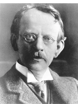
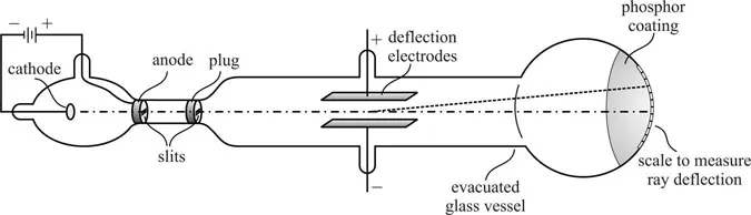
During the 1890s, many other significant experiments were being conducted with cathode ray tubes. Cathode ray tubes were built in many configurations, but common features included a neg-atively charged electrode (cathode) and a positively charged electrode (anode) sealed in an elon-gated vacuum tube, where the far end of the tube opposite the cathode was coated with a phosphor. In 1895, through the use of a cathode ray tube, Jean Baptiste Perrin made the discovery that cathode rays were composed of negative electricity. Building on this knowledge, Joseph John Thomson (see Fig. 1.6) conducted experiments with different cathode ray tubes (CRTs) to investigate the nature of the cathode rays, where he paid special attention to evacuating the CRT. This simple precaution allowed experiments to be conducted without contaminant gases which could become ionized during the operation of the tube. A schematic of such a tube is shown in Fig. 1.7. With these tubes Thomson made three fundamental experimental observations. First, he noted that magnetic fields and electric fields could deflect the trajectory of the cathode rays.
Figure 1.6. Joseph John Thomson (1856–1940).
Figure 1.7. The cathode ray tube apparatus used by Thomson to measure the q/m ratio for cathode rays.
Further, given a known magnetic field strength, he noted that the deflection of these cathode rays was far greater than observed for positively charged hydrogen gas. Finally, by comparing the results of the cathode rays and the hydrogen gas, Thomson noted that the charge-to-mass ratio was approximately 1800 times greater for the cathode rays than for hydrogen ions.2 Because he was confident no gas was inside the cathode ray tube, in 1897 Thomson correctly deduced that cathode rays were in fact negatively charged particles emitted from the cathode surface or, more correctly, emitted from the atoms composing the cathode. Although Thomson named these newly identified negative particles “corpuscles,” the eventual name given to them was electrons as suggested by George Johnston Stoney, who had actually predicted the presence of these negative particles as early as 1874.
It should be noted that Thomson’s discovery was the first recorded indication that atoms consisted of subatomic particles, thereby, earning him the 1906 Nobel Prize in Physics. Thomson’s discovery was made possible, in part, with another type of radiation detector, namely the fluorescent screen of the CRT.
The observations and findings of Becquerel caught the interests of Pierre and Marie Curie, both chemists by education (see Fig. 1.8). Marie began studying under Becquerel, and began a systematic investigation of the known elements to determine if other materials emitted energetic rays similar to those from uranium. While studying pitchblende, an uranium ore containing various oxides, thorium, and other rare earth elements, she found that thorium and thorium compounds also emitted energetic rays. Marie is generallycredited with recommending the name radioactivity3 to describe these energetic emissions. Marie quickly reported these findings, only to learn that German physicist Gerhard Schmidt had published similar findings on thorium only two months earlier. However, of genuine interest in Curie’s work was the observation that the pitchblende, after removing the uranium, was more radioactive than the uranium. The Curies thus deduced correctly that another radioactive element mus...
Table of contents
Cover
Half Title
Title Page
Copyright Page
Dedication
About the Cover Picture
Contents
Preface
About the Authors
1. Origins
2. Introduction to Nuclear Instrumentation
3. Basic Atomic and Nuclear Physics
4. Radiation Interactions
5. Sources of Radiation
6. Probability and Statistics for Radiation Counting
7. Source and Detector Effects
8. Essential Electrostatics
9. Gas-Filled Detectors: Ion Chambers
10. Gas-Filled Detectors: Proportional Counters
11. Gas-Filled Detectors: Geiger-Müller Counters
12. Review of Solid State Physics
13. Scintillation Detectors and Materials
14. Light Collection Devices
15. Basics of Semiconductor Detector Devices
16. Semiconductor Detectors
17. Slow Neutron Detectors
18. Fast Neutron Detectors
19. Luminescent and Additional Detectors
20. Radiation Measurements and Spectroscopy
21. Mitigating Background
22. Nuclear Electronics
Appendix A: Fundamental Physical Data and Conversion Factors
Appendix B: Cross Sections and Related Data
Index
Frequently asked questions
Yes, you can cancel anytime from the Subscription tab in your account settings on the Perlego website. Your subscription will stay active until the end of your current billing period. Learn how to cancel your subscription
No, books cannot be downloaded as external files, such as PDFs, for use outside of Perlego. However, you can download books within the Perlego app for offline reading on mobile or tablet. Learn how to download books offline
Perlego offers two plans: Essential and Complete
Essential is ideal for learners and professionals who enjoy exploring a wide range of subjects. Access the Essential Library with 800,000+ trusted titles and best-sellers across business, personal growth, and the humanities. Includes unlimited reading time and Standard Read Aloud voice.
Complete: Perfect for advanced learners and researchers needing full, unrestricted access. Unlock 1.5M+ books across hundreds of subjects, including academic and specialized titles. The Complete Plan also includes advanced features like Premium Read Aloud and Research Assistant.
Both plans are available with monthly, semester, or annual billing cycles.
We are an online textbook subscription service, where you can get access to an entire online library for less than the price of a single book per month. With over 1.5 million books across 990+ topics, we’ve got you covered! Learn about our mission
Look out for the read-aloud symbol on your next book to see if you can listen to it. The read-aloud tool reads text aloud for you, highlighting the text as it is being read. You can pause it, speed it up and slow it down. Learn more about Read Aloud
Yes! You can use the Perlego app on both iOS and Android devices to read anytime, anywhere — even offline. Perfect for commutes or when you’re on the go. Please note we cannot support devices running on iOS 13 and Android 7 or earlier. Learn more about using the app
Yes, you can access Radiation Detection by Douglas McGregor,J. Kenneth Shultis in PDF and/or ePUB format, as well as other popular books in Technology & Engineering & Political Freedom. We have over 1.5 million books available in our catalogue for you to explore.