The Peripheral T-Cell Lymphomas
  1. English
  2. ePUB (mobile friendly)
  3. Available on iOS & Android
eBook - ePub

About this book

THE PERIPHERAL T-CELL LYMPHOMAS

Provides a comprehensive look at Peripheral T-Cell lymphomas, including the group's unique geographic distribution, underlying genetics, and novel treatments

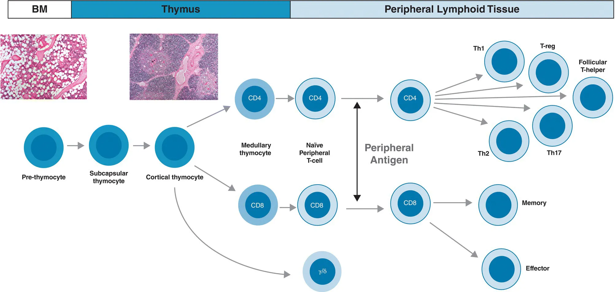
Peripheral T-Cell lymphomas (PTCL) are a diverse group of lymphoid malignancies that develop from mature T cells and natural killer (NK) cells. PTCL represent 10-15% of all cases of non-Hodgkin lymphoma in the US, and up to 20-25% of cases in South America, Asia, and other regions around the world. The role of different etiologic factors and the variation of geographic distribution makes PTCL one of the most difficult types of cancer to understand and treat.

For the first time in a single volume, The Peripheral T-Cell Lymphomas presents a comprehensive survey of this complex and rare group of blood cancers. Featuring contributions from an international team of leading authorities in the various aspects of PTCL, this authoritative text covers biology, epidemiology, classification, approved and emerging drugs, molecular genetics, and more. Detailed clinical chapters address diagnosis, prognosis, and treatment of each of the major PTCL subtypes identified in the 2018 WHO Classification of Tumors of Hematopoietic and Lymphoid Tissues. This much-needed resource:

  • Covers the biological basis, epidemiology, classification, and treatment of PTCL
  • Discusses the future of the field, including global collaboration efforts and novel approaches to PCTL
  • Explores the role of biologics in PTCL and autologous and allogeneic stem-cell transplantation
  • Offers new insights on molecular pathogenesis, innovative therapeutics, and novel drug combinations
  • Features contributions from the Chairs The T-Cell Lymphoma Forum: the world's largest meeting focused on PTCL

Reflecting the unique epidemiology and genetic diversity of the PTCL, The Peripheral T-Cell Lymphomas is an indispensable source of data, insight, and references for the medical community, particularly oncologists and hematologists in both training and practice.

Frequently asked questions

Yes, you can cancel anytime from the Subscription tab in your account settings on the Perlego website. Your subscription will stay active until the end of your current billing period. Learn how to cancel your subscription.
No, books cannot be downloaded as external files, such as PDFs, for use outside of Perlego. However, you can download books within the Perlego app for offline reading on mobile or tablet. Learn more here.
Perlego offers two plans: Essential and Complete
  • Essential is ideal for learners and professionals who enjoy exploring a wide range of subjects. Access the Essential Library with 800,000+ trusted titles and best-sellers across business, personal growth, and the humanities. Includes unlimited reading time and Standard Read Aloud voice.
  • Complete: Perfect for advanced learners and researchers needing full, unrestricted access. Unlock 1.4M+ books across hundreds of subjects, including academic and specialized titles. The Complete Plan also includes advanced features like Premium Read Aloud and Research Assistant.
Both plans are available with monthly, semester, or annual billing cycles.
We are an online textbook subscription service, where you can get access to an entire online library for less than the price of a single book per month. With over 1 million books across 1000+ topics, we’ve got you covered! Learn more here.
Look out for the read-aloud symbol on your next book to see if you can listen to it. The read-aloud tool reads text aloud for you, highlighting the text as it is being read. You can pause it, speed it up and slow it down. Learn more here.
Yes! You can use the Perlego app on both iOS or Android devices to read anytime, anywhere — even offline. Perfect for commutes or when you’re on the go.
Please note we cannot support devices running on iOS 13 and Android 7 or earlier. Learn more about using the app.
Yes, you can access The Peripheral T-Cell Lymphomas by Owen A. O'Connor, Won Seog Kim, Pier L. Zinzani, Owen A. O'Connor,Won Seog Kim,Pier L. Zinzani in PDF and/or ePUB format, as well as other popular books in Medicine & Hematology. We have over one million books available in our catalogue for you to explore.

Information

Year
2021
Print ISBN
9781119671312
eBook ISBN
9781119671367
Edition
1
Subtopic
Hematology

Part I
Biological Basis of the Peripheral T‐cell Lymphomas

1
The Fundamentals of T‐cell Lymphocyte Biology

Claudio Tripodo1,2 and Stefano A. Pileri3
1 Tumor Immunology Unit, Department of Health Sciences, University of Palermo, Palermo, Italy
2 Histopathology Unit, FIRC Institute of Molecular Oncology, Milan, Italy
3 Hematopathology Division, European Institute of Oncology, IRCCS, Milan, Italy

TAKE HOME MESSAGES

  • The natural diversity of T cells in normal immune system functions contributes – in part – to the diversity of T‐cell malignancies.
  • CD4‐positive T lymphocytes, also called T helper (Th) cells, are divided into a diverse repertoire of T lymphocytes (e.g. Th1, Th2, and Th17), in part defined by the cytokine profile they elaborate.
  • Th1 and Th2 lymphocytes can also be classified based on the expression of the transcription factors T‐bet and GATA3. These factors can be prognostic in peripheral T‐cell non‐Hodgkin lymphomas derived from these cells (with GATA3 being associated with a poor prognosis).

Introduction

The T‐cell system is conventionally regarded as an enabler of diverse compartments, which correspond to different steps of differentiation and functional subsets of mature cells taking part in the immune response in the peripheral lymphoid and non‐lymphoid tissues.
In this chapter, we give a overview of the T‐cell system, more functionally than anatomically oriented, to reflect its extreme plasticity. This plasticity is thought to lie at the heart of the diversity of T‐cell malignancies now recognized by the World Health Organization.

General View of the Differentiation and Function of T Lymphocytes

The immune system can be classified into two basic component: (i) the innate immune system, and (ii) the acquired immune system. The innate immune system is considered to be relatively agnostic to any specific antigen, and is often described as invariant. The innate immune response is the first line of defense, and typically exhibits limited specificity. Examples of innate immune response may include phagocytosis by macrophages, barriers to infection provided by the skin and tears, natural killer and mast cells, and complement‐mediated cytolysis. In contrast, the adaptive (or sometimes called acquired) immune response develops in response to specific antigen, being “custom” designed for the antigen in question. It usually occurs later in the immune response, and has the ability to recall the response to past infections. Components of a functioning acquired immune response might involve antigen‐presenting cells presenting antigen or T cells, the activation of specific T cells which would signal to B cells enlisting their engagement in the response and the production of highly specific antibody capable of binding specific antigen. T and B lymphocytes are the major types of lymphocytes found in the human body, where they can constitute 20–40% of all white blood cells, with only about 2–3% being found in the peripheral circulation, the remainder being localized to various lymphoid organs (lymph nodes, spleen, submucosal tissue). Remarkably, the total mass of lymphocytes in the body can approximate the mass of the brain and liver.
As shown in Figure 1.1 [1], T lymphocytes arise from a bone marrow precursor, which undergoes maturation and functional orientation in the thymus. Antigen‐specific T cells mature in the thymic cortex, where the elements recognizing self‐peptides and major histocompatibility antigens expressed by cortical epithelial cells and thymic nurse cells are eliminated via apoptosis. Failure to eliminate those T cells recognizing self‐peptides is thought to give rise to a host of autoimmune disorders.
image
Figure 1.1 Schematic overview of T‐cell ontogeny and differentiative trajectories.
Source: Claudio Tripodo, Stefano Pilleri.
Cortical thymocytes exhibit an immature T‐cell phenotype and express a characteristic repertoire of proteins including TdT, CD1a, CD3, CD5, and CD7. CD3 is expressed in the cytoplasm until completion of T‐cell receptor (TCR) gene rearrangement and is then exported to the cell membrane. Cortical thymocytes are initially CD4/CD8 double negative.
Medullary thymocytes exhibit a phenotype similar to that of mature T cells of the peripheral lymphoid organs with segregation of CD4 and CD8 antigens. Based on the structure of the variable portion of the TCR, T cells have been divided into two classes, including alpha/beta and gamma/delta T cells. They are both associated with the CD3 complex, which contains gamma, delta, and epsilon chains. Gamma and delta T cells usually lack expression of CD4, CD8, and CD5, although a subpopulation can expresses CD8. They represent less than 5% of normal T lymph...

Table of contents

  1. Cover
  2. Table of Contents
  3. Title Page
  4. Copyright Page
  5. Contributors
  6. About the Companion Website
  7. Part I: Biological Basis of the Peripheral T‐cell Lymphomas
  8. Part II: Epidemiology and Classification of the PTCL
  9. Part III: Discrete Clinical Subtypes of PTCL (Unique Epidemiology, Therapy and Management)
  10. Part IV: Treatment of the PTCL
  11. Part V: Future Directions
  12. Index
  13. End User License Agreement