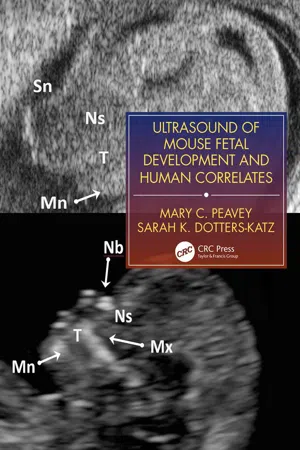
Fetal development in the mouse is routinely and increasingly utilized for advancing translational research and medical innovation for human obstetrical care. This is the first and only manual to provide necessary content on how this should be handled for accurate and effective data collection. Detailed descriptions and examples demonstrate how researchers and clinicians can use murine fetal and obstetrical data to improve future human applications in diseases such as infertility, recurrent pregnancy loss, intrauterine fetal growth restriction, placental insufficiency, and intrauterine fetal demise, as well as organ-specific developmental disease.
eBook - ePub
Ultrasound of Mouse Fetal Development and Human Correlates
- 90 pages
- English
- ePUB (mobile friendly)
- Available on iOS & Android
eBook - ePub
Ultrasound of Mouse Fetal Development and Human Correlates
About this book
Trusted by 375,005 students
Access to over 1.5 million titles for a fair monthly price.
Study more efficiently using our study tools.
Information
Topic
Medicine1 | Implantation and Embryonic Imaging Mouse until D4.5–9.5; Human 5–9 Weeks |
DATING
In the human, the first trimester marks the development of a zygote into a recognizable human fetus and is most commonly dated in relation to the first day of the last menstrual period of the mother. Consequently, the traditional dating of a human pregnancy is 2 weeks more than the actual embryological development of the pregnancy. In the mouse pregnancy, a vaginal plug is seen on the morning after copulation, and traditionally, the pregnancy is dated as this morning as being day 0.5 of pregnancy, or 0.5 days post coitus (dpc). Subsequent description of gestational dating in the human and mouse will thus be assigned as per this nomenclature.
IMAGING
In humans, during the first trimester of pregnancy, transvaginal approach provides the most accurate and reliable images of the pregnancy. In the mouse, transvaginal ultrasonography technology has yet to be developed; however the images obtained by the transabdominal approach are currently more than adequate for early gestation imaging; all mouse images were obtained with the use of the Vevo 2100 system from Fujifilm VisualSonics, as previously published (1,2). In both human and murine pregnancies, early ultrasound allows for detection of the number and location of intrauterine pregnancies, assign a gestational age and developmental of the pregnancy, and to evaluate the whether there are sonographic findings indicative of early pregnancy compromise or failure.
PREIMPLANTATION DEVELOPMENT
Similarly to human ovulation and fertilization, the mouse embryo after ovulation is transported through the fallopian tube, where fertilization with sperm occurs. The fertilized oocyte is known as the zygote, which will continue to divide into an embryo. As the embryo travels through the fallopian tube, it continues to undergo cell division; by the third day, the embryo is approximately eight cells and begins compaction. On its fifth day of growth, the human embryo has developed into a blastocyst and travels to the uterus where it begins the process of implantation and further growth. At this point, as in humans, the blastocyst consists of a discrete inner cell mass, within a spherical cavity lined by the trophectoderm cell layer. These preimplantation events cannot be ascertained via ultrasonographic methods in either the human or mouse. However, the non pregnant uterus, consisting of the myometrium and endometrium can be easily measured via sonographic methods. See Fig. 1.1.

Figure 1.1 A: The non-pregnant human uterus, with trilaminar endometrial lining pattern. B: The non pregnant mouse uterus during proestrus, appearing as an echogenic linear line in the middle of the longitudinal uterine horn.
DECIDUALIZATION AND EARLY IMPLANTATION
Changes within the endometrium in early pregnancy can be easily identified by ultrasonography, in which the endometrium appears increased in echogenicity and thickness, in both the human as well as the mouse (3–5). The decidualized endometrium is the functional layer of the edematous and thickened endometrium in which successful implantation must occur (6,7). This change in the endometrium, prior to the development of a gestational sac correlate to 4–5 weeks gestational age in humans and 3.5–6.5 dpc in the mouse.
HUMAN
The blastocyst consists of a discrete inner cell mass, within a spherical cavity lined by the trophectoderm cell layer. In humans, by the end of the third gestational week, the blastocyst begins to implant into the decidualized endometrium. In successful pregnancies, the average endometrial thickness is 17 ± 6.7 mm (8), and the distinct echogenic decidualized endometrium can be easily identified on transvaginal ultrasound, before the presence of a gestational sac. The endometrium will continue to appear thickened and echogenic from 4 to 5 weeks gestation, before the gestational sac is visible.
MOUSE
In the mouse by 3.5 dpc, the endometrium itself remains linear in appearance without discrete sites of implantation detectable; the hyperechoic endometrium is approximately 0.14–0.2 mm in thickness. However, by 4.5 dpc, the discrete locations of embryo implantation sites can be identified, as represented by a small hyperechoic dot in the middle of the expanding myometrium. By 5.5 dpc, these discrete implantation sites are easily identified and counted, allowing for the number of implantation sites as well as the spacing interval, to be determined. The average distance between each implantation site be quantified. In this animal model, at 5.5 dpc, the average normal distance between implantation sites has been determined to be 1.8–2 mm (2). At 6.5 dpc, the uterus begins to take on a distinct spherical shape around each implantation site, causing each uterine horn to resemble “a string of pearls” on ultrasound imaging. The bright areas of decidualized endometrium continue to reveal distinct endometrial implantation sites are easily identifiable; at this stage, the average spacing between each implantation site is 2.99 ± 0.3 mm (2). See Figs. 1.2–1.5.

Figure 1.2 A: The human endometrium during very early pregnancy, which is thickened at a gestational age of 4 weeks and 3 days. B: The mouse pregnancy at 3.5 dpc, with the slight appearance of three rounded implantation sites in the longitudinal uterine horn. C: The same mouse pregnancy at 4.5 dpc, with the bright echogenic implantation sites now easily visible and loss of echogenicity between implantation sites.

Figure 1.3 The early pregnant mouse pregnancy, seen here at 4.5 dpc, with a bright, echogenic sphere in the center of the expanding uterine horn. The maternal bladder is seen to the right.

Figure 1.4 A: The mouse pregnancy at 5.5 dpc, with four implantation sites seen in the right horn and the most proximal implantation site on the left horn. B: The mouse pregnancy at 5.5 dpc, with the distance between implantation sites measurable.

Figure 1.5 A: The mouse pregnancy at 6.5 dpc; three implantation sites are now easily visualized as distinct sites, within one uterine horn. B: The mouse pregnancy at 6.5 dpc; the distance between each implantation site is easily quantified, as shown measured.
DEVELOPMENT OF GESTATIONAL SAC
The first reliable sonographic evidence of an early intrauterine pregnancy in both the human and mouse is the presence of a gestational sac (9,10). A gestational sac is identified as a small, round, anechoic fluid collection surrounded by the hyperechoic rim of endometrial tissue. In both mice and in humans the mean sac diameter (MSD) can be obtained by measuring the diameter of the sac in each of three dimensions and then averaging the measurements (11). The gestational sac, is first visible at the fifth week of gestation in the human and at 7.5 dpc in the mouse pregnancy.
HUMAN
In humans, the position of the gestational sac should be in the mid to upper uterus. The shape should be round and fairly spherical in its dimensions. In humans, as early as the fifth week of pregnancy, a yolk sac within the gestational sac is the first sonographic confirmation of a developing intrauterine pregnancy. A pseudogestational sac can sometimes be seen in the instance of an ectopic or extra-uterine pregnancy, but would not contain a yolk sac.
MOUSE
In the mouse, a gestational sac should form at each implantation site, which should remain roughly equally distributed throughout each horn, both proximally and distally. Similar to human development, the gestational sac is visualized as a small, round, anechoic area in the site of the implantation area. The appearance of a gestational sac reliably occurs between 6.5 and 7.5 dpc. The absence of a gestational sac on 7.5 dpc confirms an abnormal pregnancy that will not progress into a viable pregnancy. See Figs. 1.6 and 1.7.

Figure 1.6 A: A human pregnancy 5 weeks and 3 days, with a normal appearing yolk sac within the gestational sac. B: A mouse pregnancy 7.5 dpc, with a gestational sac and likely the beginning growth of the fetus. In this instance, the measurements of the gestational sac are 1.1 × 0.7 × 0.8 mm, with a mean sac diameter (MSD) of 0.87 mm. A small echogenic area, likely a small fetal pole, is measured at 0.5 mm.

Figure 1.7 A: A human pregnancy 5 weeks and 6 days; with a normal appearing yolk sac within ...
Table of contents
- Cover
- Half Title
- Title Page
- Copyright Page
- Contents
- Acknowledgements
- Foreword
- Preface
- Chapter 1 Implantation and Embryonic Imaging: Mouse until D4.5–9.5; Human 5–9 Weeks
- Chapter 2 Early Organogenesis and First Trimester: Mouse D10.5–12.5; Human 9–12 Weeks
- Chapter 3 Mid-Gestation and Second Trimester: Mouse D13.5–15.5; Human 13–27 Weeks
- Chapter 4 Central Nervous System and Facial Development: Mouse D13.5–15.5; Human 13–27 Weeks
- Chapter 5 Late-Gestation and Third Trimester: Mouse D16.5–18.5; Human 28–40 Weeks
- Chapter 6 Placenta Throughout Gestation
- Index
Frequently asked questions
Yes, you can cancel anytime from the Subscription tab in your account settings on the Perlego website. Your subscription will stay active until the end of your current billing period. Learn how to cancel your subscription
No, books cannot be downloaded as external files, such as PDFs, for use outside of Perlego. However, you can download books within the Perlego app for offline reading on mobile or tablet. Learn how to download books offline
Perlego offers two plans: Essential and Complete
- Essential is ideal for learners and professionals who enjoy exploring a wide range of subjects. Access the Essential Library with 800,000+ trusted titles and best-sellers across business, personal growth, and the humanities. Includes unlimited reading time and Standard Read Aloud voice.
- Complete: Perfect for advanced learners and researchers needing full, unrestricted access. Unlock 1.5M+ books across hundreds of subjects, including academic and specialized titles. The Complete Plan also includes advanced features like Premium Read Aloud and Research Assistant.
We are an online textbook subscription service, where you can get access to an entire online library for less than the price of a single book per month. With over 1.5 million books across 990+ topics, we’ve got you covered! Learn about our mission
Look out for the read-aloud symbol on your next book to see if you can listen to it. The read-aloud tool reads text aloud for you, highlighting the text as it is being read. You can pause it, speed it up and slow it down. Learn more about Read Aloud
Yes! You can use the Perlego app on both iOS and Android devices to read anytime, anywhere — even offline. Perfect for commutes or when you’re on the go.
Please note we cannot support devices running on iOS 13 and Android 7 or earlier. Learn more about using the app
Please note we cannot support devices running on iOS 13 and Android 7 or earlier. Learn more about using the app
Yes, you can access Ultrasound of Mouse Fetal Development and Human Correlates by Mary C. Peavey,Sarah K. Dotters-Katz in PDF and/or ePUB format, as well as other popular books in Medicine & Gynecology, Obstetrics & Midwifery. We have over 1.5 million books available in our catalogue for you to explore.