Pediatric Dermatology in Skin of Color
eBook - ePub

Pediatric Dermatology in Skin of Color

A Practical Guide

  1. 320 pages
  2. English
  3. ePUB (mobile friendly)
  4. Available on iOS & Android
eBook - ePub

Pediatric Dermatology in Skin of Color

A Practical Guide

About this book

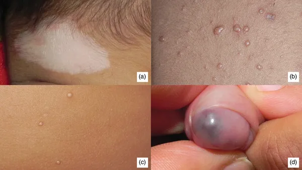
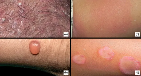
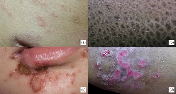
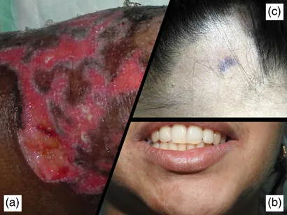
This book focusses on the clinical aspects and management of pediatric skin disorders, especially seen in darker skin types. It includes unique conditions that the authors have encountered in their lifetime with their independent observations and approach to management. Original high-quality images are used to illustrate most dermatoses described in the book enabling a strong visual impression of the discussed diseases. It hopes to provide readers with a blend of evidence and experience based pediatric dermatology. This book aims to be a hands-on manual that can be referred to during a busy practice as it discusses the practical approach to dermatoses.

Key Features


  • Focusses on darker skin types.


  • Examines unusual presentations with detailed clinical features.


  • Discusses the ways to differentiate between similar-appearing diseases.


  • Explores approaches to therapy, especially in resource-poor settings.


  • Covers topics with high quality illustrations.

Frequently asked questions

Yes, you can cancel anytime from the Subscription tab in your account settings on the Perlego website. Your subscription will stay active until the end of your current billing period. Learn how to cancel your subscription.
At the moment all of our mobile-responsive ePub books are available to download via the app. Most of our PDFs are also available to download and we're working on making the final remaining ones downloadable now. Learn more here.
Perlego offers two plans: Essential and Complete
  • Essential is ideal for learners and professionals who enjoy exploring a wide range of subjects. Access the Essential Library with 800,000+ trusted titles and best-sellers across business, personal growth, and the humanities. Includes unlimited reading time and Standard Read Aloud voice.
  • Complete: Perfect for advanced learners and researchers needing full, unrestricted access. Unlock 1.4M+ books across hundreds of subjects, including academic and specialized titles. The Complete Plan also includes advanced features like Premium Read Aloud and Research Assistant.
Both plans are available with monthly, semester, or annual billing cycles.
We are an online textbook subscription service, where you can get access to an entire online library for less than the price of a single book per month. With over 1 million books across 1000+ topics, we’ve got you covered! Learn more here.
Look out for the read-aloud symbol on your next book to see if you can listen to it. The read-aloud tool reads text aloud for you, highlighting the text as it is being read. You can pause it, speed it up and slow it down. Learn more here.
Yes! You can use the Perlego app on both iOS or Android devices to read anytime, anywhere — even offline. Perfect for commutes or when you’re on the go.
Please note we cannot support devices running on iOS 13 and Android 7 or earlier. Learn more about using the app.
Yes, you can access Pediatric Dermatology in Skin of Color by Manish K Shah,Preeti K Sheth in PDF and/or ePUB format, as well as other popular books in Medicine & Dermatology. We have over one million books available in our catalogue for you to explore.

Information

Publisher
CRC Press
Year
2021
Print ISBN
9781138486102
eBook ISBN
9781351046572
Edition
1
Subtopic
Dermatology

1

PRINCIPLES OF DIAGNOSIS

Lesionology

Lesions can be classified as primary (appear de novo) and secondary (develop due to a change in an already existing lesion).

Primary lesions

  1. Macule: A circumscribed alteration in the color or texture of the skin that is not elevated or depressed below the surface of the skin.
It may be
  1. Hypopigmented, e.g., pityriasis alba, pityriasis versicolor
  2. Hyperpigmented, e.g., lentigines, freckles
  3. Depigmented, e.g., vitiligo (Figure 1.1a)
    FIGURE 1.1
    FIGURE 1.1(a) Depigmented macule of vitiligo. (b) Flesh-colored papule of molluscum contagiosum.(c) Clear fluid-filled vesicles of varicella. (d) Blue vascular nodule on the penis.
  4. Erythematous, e.g., exanthems, drug rash
  5. Purpuric, e.g., thrombocytopenic purpura
  1. Papule: A circumscribed palpable elevation of the skin less than 0.5 cm in diameter.
It may be
  1. White, e.g., milium
  2. Flesh-colored, e.g., molluscum contagiosum (Figure 1.1b)
  3. Red, e.g., eczema
  4. Yellowish, e.g., xanthoma
  5. Yellowish brown, e.g., lupus vulgaris
  6. Black, e.g., melanoma
  1. Nodule: A solid mass in the skin that is either seen as an elevation or can be palpated. It is more than 0.5 cm but less than 2 cm in size (Figure 1.1d).
  • E.g.: lupus vulgaris, erythema nodosum
  • They may be soft, e.g., neurofibroma.
  • They may be hard, e.g., calcinosis cutis.
  1. Tumors: Soft or firm masses more than 2 cm in size, which can be inflammatory or noninflammatory, malignant or nonmalignant.
  2. Vesicle: Visible accumulation of fluid beneath the skin, less than 0.5 cm in size. The fluid may be
    1. Clear, e.g., chicken pox, herpes simplex (Figure 1.1c)
    2. Hemorrhagic, e.g., herpes zoster
    3. Cloudy, e.g., impetigo
  3. Pustule: Visible accumulation of free pus beneath the skin, measuring less than 0.5 cm in diameter. This may be
    1. Follicular, e.g., folliculitis (Figure 1.2a)
      FIGURE 1.2
      FIGURE 1.2(a) Pustules on the scalp. (b) Single bulla. (c) Urticarial wheal. (d) Plaque of psoriasis.
    2. Nonfollicular, e.g., impetigo, candidiasis
  4. Bulla: Visible accumulation of fluid beneath the skin, more than 0.5 cm in size. The fluid may be clear, hemorrhagic, or purulent.
Bullae may be tense, e.g., bullous pemphigoid (Figure 1.2b), or they may be flaccid, e.g., pemphigus.
  1. Wheal: Evanescent, edematous, erythematous or pale, flat elevation of various sizes, e.g., urticaria, urticarial vasculitis (Figure 1.2c).
  2. Patch: Alteration in skin color or texture measuring more than 0.5 cm, that is not elevated or depressed. One can argue that a patch is a large macule and need not be classified separately. Examples of patches include:
    1. Hypopigmented, e.g., leprosy
    2. Hyperpigmented, e.g., fixed drug eruption
  3. Plaque: Circumscribed elevated area of the skin more than 1 cm in size such that the horizontal dimensions are more than the vertical dimension of the lesion.
Plaques can be formed by coalescence of papules, e.g., psoriasis, or nodules, e.g., granuloma annulare. These are elevated plaques (Figure 1.2d).
Plaques of morphea are depressed.

Secondary lesions

  1. Scale: Abnormal accumulation or disordered shedding of the stratum corneum, which appears as visible flakes on the skin.
Types of scales are seen:
  1. Collarette scales: fine, peripherally attached and centrally detached scale at the edges of inflammatory lesions, i.e., pityriasis rosea (Figure 1.3a).
    FIGURE 1.3
    FIGURE 1.3(a) Collarette scaling in pityriasis rosea. (b) Thick fish-like scales in X-linkedichthyosis. (c) Honey-colored crusts in impetigo. (d) Excoriations in a case of atopicdermatitis.
  2. Branny scales, e.g., pityriasis versicolor, pityriasis alba.
  3. Ichthyotic scales: large brown polygonal scales (like on the body of a fish), e.g., ichthyosis vulgaris (Figure 1.3b).
  4. Silvery, micaceous (mica-like that looks sparkling), thick scales, e.g., psoriasis.
  1. Crust: Dried up exudates consisting of pus, serum, or dead inflammatory cells or just dried serous fluid. Sometimes dried blood.
Types:
  1. Dry, golden-yellow, soft, friable, and superficial crust: impetigo contagiosa (Figure 1.3c)
  2. Yellow, greasy crust: favus
  3. Thick, hard, and tough crust: third-degree burns
  4. Lamellated, elevated, brown crust: syphilis
  1. Erosion: Loss of epidermis, mostly after rupture of vesicles. There is no dermal damage. Healing occurs without or with minimal scarring.
  2. Excoriation: Abrasion by mechanical means, which usually involves only the epidermis. This is usually linear and is a sign of scratching. Excoriations are seen in scabies, dermatitis herpetiformis, and eczemas (Figure 1.3d).
  3. Ulcer: Breach in the continuity of skin or mucous membrane resulting from loss of dermis as well as epidermis, with slow molecular death. Ulcers always heal with scarring. Examples are traumatic ulcers and neuropathic(trophic) ulcers (Figure 1.4a).
    FIGURE 1.4
    FIGURE 1.4(a) Ulcer of pyoderma gangrenosum. (b) Atrophy of right mandibular region in morphea.(c) Lichenification in a patch of alopecia areata.
  4. Atrophy: Secondary change in the skin characterized by loss of normal ...

Table of contents

  1. Cover
  2. Half Title
  3. Title Page
  4. Copyright Page
  5. Dedication
  6. Contents
  7. Preface
  8. Acknowledgments
  9. Authors
  10. 1. Principles of Diagnosis
  11. 2. Neonatal Dermatology
  12. 3. Papulosquamous Disorders
  13. 4. Eczema and Dermatitis
  14. 5. Disorders of Cornification
  15. 6. Vesiculobullous Disorders
  16. 7. Nodules and Tumors
  17. 8. Disorders of Pigmentation
  18. 9. Infections and Infestations
  19. 10. Collagen Vascular Diseases
  20. 11. Urticaria and Erythemas
  21. 12. Drug Eruptions
  22. 13. Vascular Abnormalities
  23. 14. Cutaneous Nevi
  24. 15. Genodermatoses
  25. 16. Hair and Nail Abnormalities
  26. 17. Oral Cavity and Tongue
  27. 18. Systemic Diseases
  28. 19. Psychocutaneous Diseases
  29. 20. Miscellaneous Conditions
  30. Index