- 400 pages
- English
- ePUB (mobile friendly)
- Available on iOS & Android
eBook - ePub
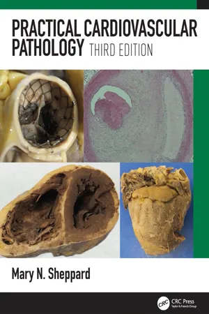
Practical Cardiovascular Pathology
About this book
In the last decade cardiac pathology has undergone a revolution, particularly in the fields of genetics and imaging. Practical Cardiac Pathology 3e is a combined atlas and text that is designed to assist pathologists in identifting the range of cardiovascular conditions that are found in both diagnostic and autopsy work.
Tools to learn more effectively

Saving Books

Keyword Search

Annotating Text

Listen to it instead
Information
Chapter One
Autopsy Cardiac Examination
Inspect heart in situ in the chest
Note fibrous outer pericardium and any fluid within
Normal – 30 ml clear serous fluid
Acute accumulation – 300 ml
Chronic – 1000 ml plus
Remove heart by cutting across great vessels and veins. Leave proximal 30 mm of aorta and pulmonary artery. Also, leave 10 mm of superior vena cava and cut inferior vena cava close to diaphragm
Inspect epicardial external surface of heart
Measure heart diameter at AV junction posteriorly
Measure longitudinal length of ventricles from crux, where right coronary artery (RCA) bends down into posterior interventricular groove down to the apex. If circumflex is dominant, use this
Cut coronary arteries and branches transversely at 2 mm intervals down to the apex on right and left sides
Cut ventricles transversely up to tips of papillary muscles and describe any changes. Remove post-mortem blood clot before weighing the heart
Measure thickness of anterior, lateral and posterior RV, interventricular septum, anterior, lateral and posterior left ventricle at midventricular level
Cut into both atria and inspect both AV valves from above
Make lateral cuts between atria and ventricles, including AV valves
Inspect ventricles and valves. Measure atrioventricular valve circumferences
Open up into right and left ventricular outflow tracts and inspect valves and origin of the coronary arteries. Measure ventriculoarterial valve circumference
Inspect aorta and pulmonary arteries. Measure circumference 10 mm above valves
Autopsy practice is changing in the modern world. There has been a marked reduction in the number of autopsies performed in the UK and elsewhere. Expertise is thus being reduced and detailed anatomical knowledge with variations of the normal organs is becoming limited. Vascular diseases of the brain and heart account for the majority of deaths in developed countries and in-depth knowledge of both these organs is essential.
The excised heart displayed grossly, recorded photographically, measured carefully and studied histologically remains the gold standard against which antemortem clinical findings are measured. Exact measurements are needed to confirm cardiac atrial dilatation, ventricular hypertrophy or dilatation, critical valve stenosis, valve regurgitation and aortic dilatation, as well as coronary artery narrowing.
When carrying out a cardiac autopsy, particularly in someone who has died suddenly with no previous disease and the most likely cause of death is in the heart, one has to look at the heart very carefully. One must take well-chosen histological sections and collect material for microbiology and particularly genetic studies. Forethought and preparation on the part of the pathologist is essential in the approach to the cardiac autopsy.
In the UK and the US, most autopsies are performed at the request of the coroner or medical examiner, with few in-hospital autopsies being carried out. Most autopsies ordered by the medical examiner/coroner are in patients where no death certificate can be issued. The majority are those who had not been to a doctor recently and did not have a life-threatening disease or were not expected to die. Of 500 000 deaths in England, 23% result in an autopsy, the majority being coroners’ cases, amounting to 94 000 per year.1 Most are carried out by a pathologist working in a general hospital or a forensic pathologist. Retention of tissues and organs from a coronial autopsy without relatives’ consent is permissible under Coroner’s Rule 9, to confirm the cause of death. If tissue is retained beyond the coroner’s investigation, the family must consent to this under the Human Tissue Act of 2004.2 I believe that pathologists must prepare in advance, especially with young, sudden deaths, and must be proactive in making a case to retain heart tissue in order to provide as accurate a cause of death as possible. They must communicate via a specifically trained coroner’s officer, who talks with the family before undertaking the post mortem to prepare them for the possibility of retention of the heart and other tissues. The help of a well-trained coroner’s officer who links with the family directly is essential in these situations for obtaining appropriate consent.
There are well established and published guidelines for pathologists investigating sudden death in both the UK and Europe.3,4
All autopsy practitioners should be able to perform a basic examination of the heart and its connecting vasculature – akin to the minimum dataset for a cancer report. Minimal information, with limited formulaic descriptions of the heart with no measurements, is unacceptable. There is a balance to be derived between the majority of autopsy cardiac cases recognized to be routine and those requiring greater consideration.
The pathologist must approach the heart armed with information about the patient’s background and the circumstances of death. Information from the general practitioner, family and witnesses is usually obtained from the coroner’s office or the medical examiner’s office, particularly in cases of unexplained sudden death. Communication with relevant cardiac centres and access to clinical records are also essential when the patient has previous cardiac interventions or surgery, which will be dealt with in Chapter 9.
Consideration of family consent is essential before the autopsy and critical if considering retaining the heart and other tissues. Specialist investigation, including culture⁄transport media for toxicology, microbiology and DNA extraction, should be taken into account prior to the commencement of the dissection in order to optimize sampling. I believe a pathologist approaching a post mortem in circumstances where the dead person has had no medical history, is failing in their duty if they do not approach the case as paediatric pathologists approach a sudden infant death, where there are established protocols to be followed.5
Digital photography is a quick, useful adjunct to autopsy diagnosis and camera facilities should be available in every mortuary. Digital images of mid–low ventricular transverse sections and other views of the heart are helpful as a permanent record and for referral when the heart cannot be retained. In sudden cardiac death, organ retention and referral should be regarded as the ‘gold standard’, with cardiac examination, tissue block and sectioning with staining being done immediately and turnaround of cases being complete within 2 weeks also with toxicology. Families can be reassured that the bulk (usually more than 90% of the cardiac tissues) can be reunited with the body in such circumstances, once the examination is complete.
Approach to the Heart in the Chest
The heart lies in the middle of the inferior mediastinum, mainly to the left of the midline behind the second to the sixth costal cartilage, with the left edge extending to the midclavicular line (Fig. 1.1). On each side, the heart abuts the lungs and the pleural cavity overlies the right side of the heart as far ...
Table of contents
- Cover
- Half Title
- Title Page
- Copyright Page
- Dedication
- Contents
- Preface
- Useful Abbreviations
- Chapter 1: Autopsy cardiac examination
- Chapter 2: The coronary arteries: Atherosclerosis and ischaemic heart disease
- Chapter 3: Valve disease
- Chapter 4: Infective endocarditis
- Chapter 5: Cardiac hypertrophy, heart failure and cardiomyopathy
- Chapter 6: Myocarditis
- Chapter 7: Cardiac tumours
- Chapter 8: Diseases of the aorta
- Chapter 9: Deaths following cardiac surgery and invasive interventions
- Chapter 10: Investigation of sudden cardiac death
- Chapter 11: Arteropathies, microcirculation and vasculitis
- Chapter 12: Pericardium
- Index
Frequently asked questions
Yes, you can cancel anytime from the Subscription tab in your account settings on the Perlego website. Your subscription will stay active until the end of your current billing period. Learn how to cancel your subscription
No, books cannot be downloaded as external files, such as PDFs, for use outside of Perlego. However, you can download books within the Perlego app for offline reading on mobile or tablet. Learn how to download books offline
Perlego offers two plans: Essential and Complete
- Essential is ideal for learners and professionals who enjoy exploring a wide range of subjects. Access the Essential Library with 800,000+ trusted titles and best-sellers across business, personal growth, and the humanities. Includes unlimited reading time and Standard Read Aloud voice.
- Complete: Perfect for advanced learners and researchers needing full, unrestricted access. Unlock 1.4M+ books across hundreds of subjects, including academic and specialized titles. The Complete Plan also includes advanced features like Premium Read Aloud and Research Assistant.
We are an online textbook subscription service, where you can get access to an entire online library for less than the price of a single book per month. With over 1 million books across 990+ topics, we’ve got you covered! Learn about our mission
Look out for the read-aloud symbol on your next book to see if you can listen to it. The read-aloud tool reads text aloud for you, highlighting the text as it is being read. You can pause it, speed it up and slow it down. Learn more about Read Aloud
Yes! You can use the Perlego app on both iOS and Android devices to read anytime, anywhere — even offline. Perfect for commutes or when you’re on the go.
Please note we cannot support devices running on iOS 13 and Android 7 or earlier. Learn more about using the app
Please note we cannot support devices running on iOS 13 and Android 7 or earlier. Learn more about using the app
Yes, you can access Practical Cardiovascular Pathology by Mary N. Sheppard in PDF and/or ePUB format, as well as other popular books in Medicine & Cardiology. We have over one million books available in our catalogue for you to explore.