Human Microanatomy is a comprehensive histology text that analyzes human structure and function from the subcellular to organ level of organization. In addition to emphasizing medically relevant information, each chapter considers developmental and evolutionary aspects of microanatomy while also using celebrity medical histories to help provide real-world context for accompanying descriptions of normal histology. The book is richly illustrated with over 1400 full-color micrographs and drawings assembled into cohesive groupings with detailed captions to help elucidate key histological concepts. Text illustrations are further supplemented by hundreds of other light and electron micrographs available in a free digital atlas covering a broad spectrum of microanatomy. Each text chapter also includes a preview, pictorial summary, and self-study quiz to highlight and review essential elements of histology. By incorporating features like medical histories, biological correlates, and various study aids, Human Microanatomy provides an appealing and informative treatment of histology for readers who are interested in the structural bases of cell, tissue, and organ functioning.
KEY FEATURES:
Uses celebrity medical histories to help provide context for descriptions of normal histology
Supplements medically relevant information with developmental and evolutionary correlates of microanatomy
Contains 1400+ full-color micrographs and drawings that illustrate a wide range of histological features
Offers free access to an ancillary online atlas with hundreds of additional light and electron micrographs
Includes helpful study aids such as chapter previews, pictorial summaries, and self-study quizzes
Presents a novel and comprehensive account of the structure and function of human cells, tissues, and organs
Trusted by 375,005 students
Access to over 1.5 million titles for a fair monthly price.
PART ONECELL AND TISSUE HISTOLOGY—OVERVIEW OF HISTOLOGY, CELL STRUCTURE, AND TISSUES
1Introduction to Human Microanatomy and its Associated Techniques
2Primer of Cellular Ultrastructure
3Overview of Embryology and the Four Basic Tissue Typeswith Emphasis on Epithelium and Connective Tissue Proper
4Cartilage and Bone
5Adipose Tissue
CHAPTER1Introduction to Human Microanatomy and its Associated Techniques
DOI: 10.1201/9780429353307-2
PREVIEW
1.1 Human microanatomy and the general organization of this textbook
1.2 Cells, size scales, and limits of resolution in light microscopy (LM) vs. electron microscopy (EM)
1.3 Imaging live whole mounts by LM and preparing fixed sections for LM and TEM
1.4 Staining sections, histological artifacts, and interpreting microanatomical images
1.5 Identifying specific components in histological samples: polarization microscopy, autoradiography, and indirect immunofluorescence
1.6 Methods for reconstructing three-dimensional morphology from the subcellular to organ level of organization
1.7 Medical applications of microanatomical analyses
1.8 Summary and self-study questions
1.1HUMAN MICROANATOMY AND THE GENERAL ORGANIZATION OF THIS TEXTBOOK
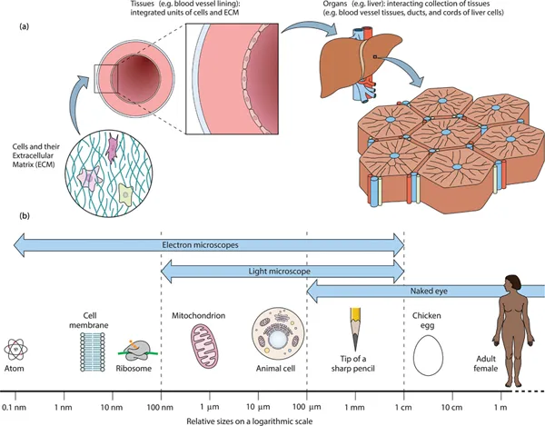
During animal evolution, individual cells and their surrounding networks of molecules that constitute the extracellular matrix (ECM) have aggregated to form functionally integrated assemblages, called tissues (Figure 1.1a). Tissues, in turn, join together to create organs (Figure 1.1a), each of which comprises a discrete collection of interacting tissues with differing functional properties and developmental origins (Chapter 3). The structure and function of cells, tissues, and organs are the focus of histology (= Greek: “histo” [woven, web, tissue] + “logos” [study of]). Alternatively, because histological studies often rely on microscopes to analyze anatomical components, histology is also known as microscopic anatomy or microanatomy.
Figure1.1Introduction to microanatomy and its size scales. (a) Microanatomy (=histology) analyzes the functional morphology of (i) cells, (ii) tissues (=integrated assemblages of cells plus their extracellular matrix [ECM]), and (iii) organs (=discrete collections of interacting tissues). (b) Some biological structures with conserved size ranges include (i) cells (typically 10–50 µm in diameter), (ii) mitochondria (∼0.5–1 µm wide), (iii) ribosomes (∼25 nm in diameter), and (iv) cell membranes (=plasma membranes) (7 nm thick).
This book covers the fundamentals of human microanatomy from a biologically oriented point of view that incorporates developmental and evolutionary perspectives into its descriptions of functional morphology. Although medically relevant information is routinely presented, detailed depictions of pathological microanatomy are seldom included. Instead, this text aims to provide a concise account of the normal structure and function of cells, tissues, and organs. Accordingly, overviews of cell and tissue biology presented in Chapters 2 and 3 focus on essential background information needed for subsequent chapters covering specific cells and tissues in normally functioning organs. To help provide orientation for descriptions of organ-level microanatomy, many of the included micrographs have stitched together overlapping regions to generate wide-field panoramic views of whole organs or large portions of organs. Thus, owing to size limitations or the unavailability of suitable human material, some images depict the microanatomy of smaller animals such as mice, which nevertheless illustrate histological features resembling those of humans. Similarly, experimental analyses of tissue and organ functioning that are highlighted throughout the text often involve animal models that can provide key insights into how the human body works.
In addition to conventional presentations of microanatomy, short medical histories are also included here as a way of lending “human faces” to histology. For such histories, 80 deceased individuals ranging from world-renowned icons to far less-famous figures have been selected in order to assemble a diverse collection of celebrities. Each of these accounts contains non-medically related information that helps flesh out its celebrity beyond simply being the bearer of a specific pathology, and by linking medical conditions to people from varied walks of life, this ancillary material is meant to provide broad context for accompanying descriptions of normal histology.
The rest of this chapter focuses on size scales, limits of resolution, and basic techniques used in microanatomy. In particular, two main methods traditionally employed in microanatomical studies—light microscopy (LM) and electron microscopy (EM)—are described while also summarizing some of the more modern ways of analyzing cell, tissue, and organ histology. Such introductory material is not intended to present in-depth explanations of the highlighted techniques but rather to offer some practical insights into the capabilities and limitations of these methods that can help with interpretations of microanatomical images included throughout the text.
1.2CELLS, SIZE SCALES, AND LIMITS OF RESOLUTION IN LIGHT MICROSCOPY (LM) VS. ELECTRON MICROSCOPY (EM)
Recent estimates indicate that the adult human body is composed of ∼30 trillion cells plus a similar number of symbiotic bacteria and other microbes that constitute the body's microbiome. Although there is no universally accepted definition of what comprises distinct cell types, a figure of ∼200 kinds of human cells is often quoted, with red blood cells accounting for the vast majority of human cells, followed distantly by such examples as bone marrow cells, endothelial cells of blood vessels, and neural cells (Table 1.1).
TABLE1.1 HUMAN CELL TOTALS
Cell type
Estimated % of ∼30 trillion cells in adult human
Erythrocytes
84
Platelets
4.9
Bone marrow cells
2.5
Vascular endothelial cells
2.1
Lymphocytes
1.5
Hepatocytes
0.8
Neurons and glia
0.6
Epidermal cells
0.5
All other cells
∼3
Source: Data from Sender, R et al. (2016) Revised estimates for the number of human and bacteria cells in the body. PLoS ONE Biology 14: doi:http://dx.doi.org/10.1371/journal.pbio.1002533, reproduced under a creative commons license.
In spite of such diversity, human cells are fairly uniform in size, with most of their diameters measuring ∼10–50 µm. Some cells such as neurons in the sciatic nerve can extend narrow axonal projections up to a meter in length. However, surface-to-volume ratios required for efficient diffusion-based processes constrain the thickness of cells. This, in turn, limits typical cellular widths to ∼100–120 µm, which is the size of fully grown human eggs and large versions of dorsal root ganglion cells near the spinal cord. Conversely, diameters of ∼5 µm in a few human cells, such as sperm or granular cells of the brain represent the minimum size for housing a nucleus plus some cytoplasmic organelles. For context, the 50-µm value for the upper end of typical cell sizes approximates the average width of scalp hairs in adult humans. Moreover, subcellular components with highly conserved sizes across numerous cell types include such examples as: (i) mitochondria, which are usually ∼0.5–1 µm wide, (ii) ribosomes with their ∼25-nm diameters, and (iii) cell membranes (=plasma membranes) that are routinely 7 nm thick (Figure 1.1b).
A single human hair placed on a contrasting background can be easily detected by an unaided human eye. However, if two closely positioned hairs are separated from each other by only 50 µm, human vision cannot discriminate between two hairs vs. only a single thick hair being present, because the eye cannot resolve such narrow spacing. Put in anothe...
Table of contents
Cover Page
Half Title Page
Title Page
Copyright Page
Dedication Page
Contents
Preface
Acknowledgments
Online Digital Atlas and Instructor Resources
Part I Cell and Tissue Histology–Overview of Histology, Cell Structure, and Tissues
Part II Tissue and Organ Histology– Blood, Circulatory and Lymphatic Systems, Muscles, Nervous System, and Skin
Part III Organ Histology—Digestive, Respiratory, and Endocrine Systems
Part IV Organ Histology– Urinary System, Reproductive Systems, and Sensory Organs
Figure Citations for Medical Histories
Index
Frequently asked questions
Yes, you can cancel anytime from the Subscription tab in your account settings on the Perlego website. Your subscription will stay active until the end of your current billing period. Learn how to cancel your subscription
No, books cannot be downloaded as external files, such as PDFs, for use outside of Perlego. However, you can download books within the Perlego app for offline reading on mobile or tablet. Learn how to download books offline
Perlego offers two plans: Essential and Complete
Essential is ideal for learners and professionals who enjoy exploring a wide range of subjects. Access the Essential Library with 800,000+ trusted titles and best-sellers across business, personal growth, and the humanities. Includes unlimited reading time and Standard Read Aloud voice.
Complete: Perfect for advanced learners and researchers needing full, unrestricted access. Unlock 1.5M+ books across hundreds of subjects, including academic and specialized titles. The Complete Plan also includes advanced features like Premium Read Aloud and Research Assistant.
Both plans are available with monthly, semester, or annual billing cycles.
We are an online textbook subscription service, where you can get access to an entire online library for less than the price of a single book per month. With over 1.5 million books across 990+ topics, we’ve got you covered! Learn about our mission
Look out for the read-aloud symbol on your next book to see if you can listen to it. The read-aloud tool reads text aloud for you, highlighting the text as it is being read. You can pause it, speed it up and slow it down. Learn more about Read Aloud
Yes! You can use the Perlego app on both iOS and Android devices to read anytime, anywhere — even offline. Perfect for commutes or when you’re on the go. Please note we cannot support devices running on iOS 13 and Android 7 or earlier. Learn more about using the app
Yes, you can access Human Microanatomy by Stephen A. Stricker in PDF and/or ePUB format, as well as other popular books in Biological Sciences & Biology. We have over 1.5 million books available in our catalogue for you to explore.