Dermoscopy can be a useful tool to evaluate skin of color for general dermatologic diseases; however, it does require practitioners to be aware of many points of difference from patients with lighter phototypes. This highly illustrated text brings together the pioneering experience of international experts to document patients of phototypes IV to VI (from subcontinental Asian, North African, South American, to African skin).
eBook - ePub
Dermoscopy in General Dermatology for Skin of Color
- 224 pages
- English
- ePUB (mobile friendly)
- Available on iOS & Android
eBook - ePub
Dermoscopy in General Dermatology for Skin of Color
About this book
Trusted by 375,005 students
Access to over 1.5 million titles for a fair monthly price.
Study more efficiently using our study tools.
Information
Topic
MedicinePart I
Inflammatory and Infiltrative Diseases
1
Papulosquamous disorders
DOI: 10.1201/9780367816483-1
1.1 Psoriasis
1.1.1 Introduction
Psoriasis is a chronic immune-mediated, inflammatory disease mainly involving the skin, nails and joints pathogenetically related to both genetic and environmental factors.1,2 Cutaneous psoriasis is typically considered as a hyperproliferative disorder with an increased proliferation of keratinocytes resulting from a cascade of immunologic reactions driven by several cytokines.1,2 The prevalence rate among dark-skinned populations varies according to the countries, yet it is generally lower than that of Caucasians.1,2 Although psoriasis can manifest at any age, it generally presents two peaks of onset (20–30 and 50–60 years of age).1,2
1.1.2 Clinical presentation
Several clinical variants of skin psoriasis do exist, with the most common one being plaque-type psoriasis (also known as psoriasis vulgaris).1,2 This form typically presents with relatively symmetric, well-defined, brownish, violaceous or greyish plaques covered with silvery scales more commonly affecting the scalp, elbows, knees, trunk, genitalia and presacral and palmo-plantar areas (Figures 1.1A–1.6A).1,2 Pruritus may be present, especially in lesions of the scalp.1,2 Residual dyspigmentations (hyper- or hypopigmentations) after the lesions heal are quite common in skin of colour (Figures 1.7A and 1.8A).1,2
Other common forms of psoriasis include guttate psoriasis (acute onset of multiple small scaly papules often triggered by a streptococcal pharyngitis), erythrodermic psoriasis (generalized erythema and scaling affecting >90% of the body surface), genital psoriasis (Figure 1.9A), palmo-plantar or diffuse pustular psoriasis (groups of pinpoint sterile pustules with or without plaques) and inverse psoriasis (well-demarcated plaques with no visible scaling involving the skin folds).1,2 Follicular psoriasis (Figure 1.10A), psoriasis ostracea, psoriasis rupioides (Figure 1.11A), psoriasis figurata, psoriasis gyrata and annular psoriasis are examples of rare variants.1,2
Finally, nail psoriasis is quite common (30%–50% of psoriatic patients) and may manifest with several ungual changes, such as nail pitting, crumbling, Beau lines and mottled lunula in case of matrix involvement and onycholysis, salmon patches, subungual hyperkeratosis and splinter haemorrhages in case of bed involvement.1,2
1.1.3 Dermoscopy
The dermoscopic hallmarks of psoriasis are represented by diffuse white scales and uniform dotted vessels (Figure 1.1B), histologically corresponding to hyperkeratosis and tips of dilated vessels in regularly elongated dermal papillae (papillomatosis), respectively.3–5 Notably, even though the prevalence of vascular structures in dark-skinned patients is lower than that in fair-skinned subjects, psoriasis is one the few papulosquamous dermatoses in which vessels are commonly seen on dermoscopy (60%–75% of cases).6,7,8 This is because of the fact that the significant epidermal acanthosis typical of psoriasis makes the skin background lighter, thus enhancing the optical contrast with vessels (Figures 1.1B and 1.2B).6
In hyperkeratotic plaques, thick scales may cover the underlying vascular structures, yet removal of the scales may bring to light the typical vascular pattern.3–6 However, analysis of the scaling pattern (colour and distribution) still remains the main dermoscopic clue in a significant proportion of dark-skinned patients, especially in areas having a thick skin (i.e., scalp or palmo-plantar areas) (Figures 1.3B–1.5B).6 Additionally, vessels are difficult to see also in initial/thin lesions as they are characterized by a limited papillomatosis on histology.3–6 Pigmentary structures (especially focal or diffuse brown structureless areas resulting from basal layer hyperpigmentation) are also not uncommonly seen in psoriasis in ethnic skin (Figure 1.1B).6,7
Further dermoscopic findings of psoriasis include focal structureless white areas (related to epidermal acanthosis) (Figure 1.1B) as well as haemorrhagic dots/areas, erosions and broken hairs resulting from scratching (Figure 1.6B), especially on the scalp and legs.3–6 Additionally, dermoscopy can also help the clinician diagnose psoriasis retrospectively as the typical vascular pattern may also be observed in hypopigmented postinflammatory patches (Figures 1.7B and 1.8B).3–5,8
The dermoscopic pattern of specific subtypes of psoriasis do not significantly differ from each other, except for the scaling amount.3–5,8 Indeed, inverse psoriasis and psoriatic balanitis (Figure 1.9B) classically do not feature scales, whereas scalp and palmo-plantar psoriasis usually reveal a thick hyperkeratotic surface (Figure 1.4B).3–5,8 Nevertheless, there are some clinical forms of psoriasis, which are not so uncommon in dark skin, that may show peculiar dermoscopic findings, such as follicular psoriasis, characterized by irregularly distributed follicular plugs associated with white scaling (Figure 1.10B) and rupioid psoriasis, which displays cone-shaped thick scales (Figure 1.11B).8
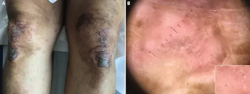
Figure 1.1 Plaque-type psoriasis in an Indian woman (A). Dermoscopy reveals diffuse white scales and un...
Table of contents
- Cover
- Half Title
- Title
- Copyright
- Dedication
- Contents
- List of contributors
- Preface
- Introduction
- Part I Inflammatory and infiltrative diseases
- Part II Infectious diseases
- Part III Hair, nail and mucosal diseases
- Index
Frequently asked questions
Yes, you can cancel anytime from the Subscription tab in your account settings on the Perlego website. Your subscription will stay active until the end of your current billing period. Learn how to cancel your subscription
No, books cannot be downloaded as external files, such as PDFs, for use outside of Perlego. However, you can download books within the Perlego app for offline reading on mobile or tablet. Learn how to download books offline
Perlego offers two plans: Essential and Complete
- Essential is ideal for learners and professionals who enjoy exploring a wide range of subjects. Access the Essential Library with 800,000+ trusted titles and best-sellers across business, personal growth, and the humanities. Includes unlimited reading time and Standard Read Aloud voice.
- Complete: Perfect for advanced learners and researchers needing full, unrestricted access. Unlock 1.5M+ books across hundreds of subjects, including academic and specialized titles. The Complete Plan also includes advanced features like Premium Read Aloud and Research Assistant.
We are an online textbook subscription service, where you can get access to an entire online library for less than the price of a single book per month. With over 1.5 million books across 990+ topics, we’ve got you covered! Learn about our mission
Look out for the read-aloud symbol on your next book to see if you can listen to it. The read-aloud tool reads text aloud for you, highlighting the text as it is being read. You can pause it, speed it up and slow it down. Learn more about Read Aloud
Yes! You can use the Perlego app on both iOS and Android devices to read anytime, anywhere — even offline. Perfect for commutes or when you’re on the go.
Please note we cannot support devices running on iOS 13 and Android 7 or earlier. Learn more about using the app
Please note we cannot support devices running on iOS 13 and Android 7 or earlier. Learn more about using the app
Yes, you can access Dermoscopy in General Dermatology for Skin of Color by Enzo Errichetti, Aimilios Lallas, Enzo Errichetti,Aimilios Lallas in PDF and/or ePUB format, as well as other popular books in Medicine & Family Medicine & General Practice. We have over 1.5 million books available in our catalogue for you to explore.