Blackwell's Five-Minute Veterinary Consult Clinical Companion
Small Animal Dentistry
Heidi B. Lobprise, Heidi B. Lobprise
- English
- ePUB (mobile friendly)
- Available on iOS & Android
Blackwell's Five-Minute Veterinary Consult Clinical Companion
Small Animal Dentistry
Heidi B. Lobprise, Heidi B. Lobprise
About This Book
The newly revised Third Edition of Blackwell's Five-Minute Veterinary Consult Clinical Companion: Small Animal Dentistry delivers an expertly edited quick-reference guide to all aspects of small animal dentistry. The book comprehensively describes new technologies and techniques as well as updated classifications and terminology. Readers willenjoy fast access to basic knowledge and detailed instructions for a wide variety of techniques in small animal dentistry.
Newer technologies, like digital radiographs and advanced images, and newer techniques, like regional blocks, are combined with the latest in treatment information to provide readers with the most logically organized reference manual in the industry. The book's companion website offers video clips and client education handouts perfect for use in a busy veterinary practice.
General practitioners will find this book to be a practical and indispensable resource. Readers will also enjoy:
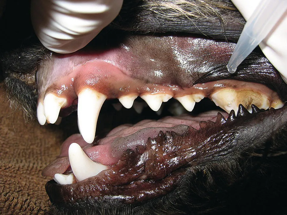
- A thorough discussion of small animal dentistry diagnostics, including oral exams and charting, periodontal probing, transillumination, and intraoral radiology and advanced imaging
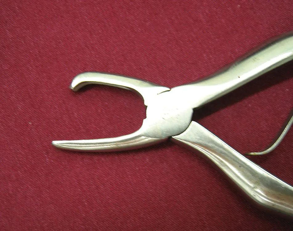
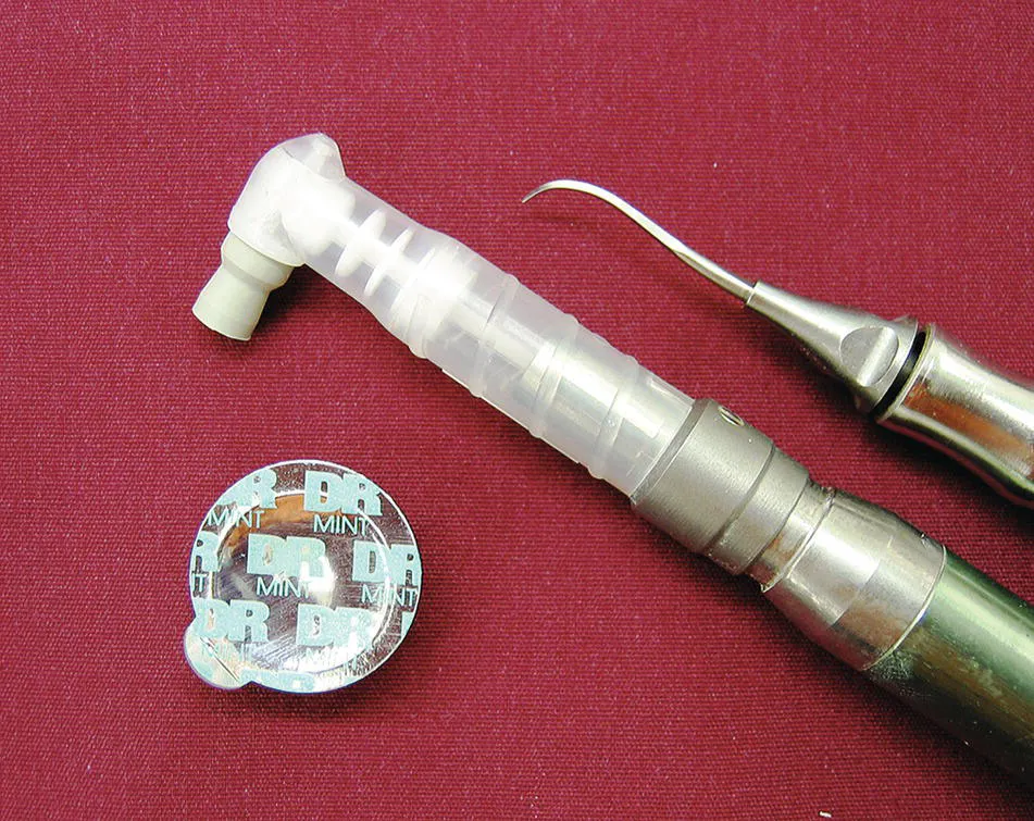
- An exploration of small animal dentistry techniques, including dental cleaning, root planing and periodontal pocket therapy, gingival flaps, extraction techniques, and oral pain management
- Analyses of a wide variety of developmental oral and dental problems, including retained deciduous teeth, dentigerous cysts, and palatal defects
Perfect for small animal general veterinary practitioners, veterinary technicians, and veterinary nurses, ; Blackwell's Five-Minute Veterinary Consult Clinical Companion: Small Animal Dentistry will also earn a place in the libraries of veterinary students who hope to improve their understanding of small animal dentistry with a quick-reference guide containing step-by-step procedures.
Frequently asked questions
Information
Section II
Techniques
Chapter 4
Complete DentalCleaning
INDICATIONS
- To remove deposits of plaque, calculus and debris from teeth.
- Another term, prophylaxis, is often used, but is less accurate as it implies prevention, which is the case only in cleaning teeth in stage 1 periodontal disease.
EQUIPMENT (see Chapter 9)
- Gloves, mask, eye protection
- Dilute chlorhexidine rinse (0.12%)
- Mouth wedge or gag
- Dental mirror
- Calculus forceps (Figure 4.1)
- Scaler (ultrasonic, sonic) (Figure 4.2)
- Hand scaler (Jacquette)
- Disclosing solution (optional)
- Slow speed polisher, prophy angle, prophy cup
- Polishing paste
- Fluoride (optional)
PROCEDURE
- General anesthesia with cuffed endotracheal tube, monitoring, and supportive care.
- Gently flush oral cavity with dilute chlorhexidine (Figure 4.3); avoid getting solution on nasal mucosa, especially in cats.
- Initial assessment to identify areas of significant disease that might require therapy, in particular those areas that were not apparent on the alert examination.
- Palatal aspect of maxillary canine – deep pockets.
- Pockets at either aspect of mandibular first molar.
- Any worn, chipped, fractured, or discolored teeth.
- Gently dislodge larger sections of calculus with calculus forceps; take care not to damage teeth (Figure 4.4).
- Use mechanized scaler (ultrasonic or sonic) to continue to remove gross deposits of calculus from crown surfaces.
- Use side of scaler tip, not end (Figure 4.5).
- Use sufficient water spray for coolant; replace scaler stack if tip overheats.
- Apply side of scaler to individual tooth for no longer than 10–12 seconds at a time; return to the tooth later if additional scaling is necessary.
- Use the sharp tip of a hand scaler (Jacquette) to remove remaining deposits of calculus in grooves (upper fourth premolar development groove); do not use tip of ultrasonic scaler there (Figure 4.6).
- Use disclosing solution or air syringe to identify any remaining deposits of calculus.
- Complete examination, probing (Figure 4.7), and intraoral radiology (see Chapters 1–3).
- Additional therapy
- Periodontal pockets (see Chapter 5).
- Extractions as needed (see Chapters 6 and 7).