1
Anatomy of the thorax
Note to reader: The main anatomical information for this chapter comes from the 40th edition of Gray’s Anatomy (Standring 2008). When content comes from an alternate source it is referenced accordingly.
The thorax contributes approximately 20 per cent to the overall length of the body compared to 12 per cent from the lumbar spine and 8 per cent from the cervical spine. While the anatomy of the thorax is highly variable between individuals, there are general similarities that are fundamental to understanding the biomechanics, assessment, and treatment of this large part of the trunk and body. There are four regions of the thorax, differentiated by the anatomy and consequential biomechanics: the vertebromanubrial, vertebrosternal, vertebrochondral, and thoracolumbar (Fig. 1.1A & B). Except for the thoracolumbar, each region is comprised of a variable number of thoracic rings (Fig. 1.2). A thoracic ring (Lee D 1994) is derived from one embryological somite which divides into a sclerotome, myotome and dermatome, and consists of the following:
Figure 1.1
(A) Three regions of the thorax are evident from the anterior aspect; the vertebromanubrial (upper thoracic rings 1 and 2), the vertebrosternal (middle thoracic rings 3 to 6) and the vertebrochondral (lower thoracic rings 7 to 10). (B) The fourth region, the thoracolumbar, does not contain ‘rings’ since the 11th and 12th ribs do not attach to the sternum.
Reproduced from Essential Anatomy 5 3D4Medical with permission.
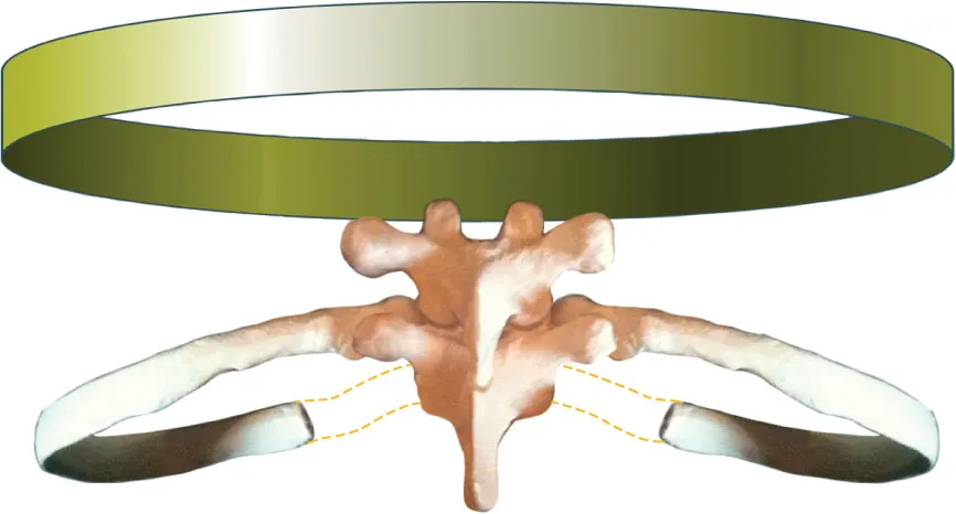
Figure 1.2
A thoracic ring as defined by Lee (Lee D 1994) consisting of the left and right ribs of the same number, the inferior half of the top vertebra and the superior half of the bottom vertebra, the left and right costocartilages, and the manubrium or sternum. There are 13 joints in each typical thoracic ring.
• Left and right ribs which are derived from the costal element of two adjacent sclerotomes (one half from each).
• Adjacent thoracic vertebrae and the intervening intervertebral discs. Each thoracic vertebra develops from two adjacent sclerotomes; therefore, the proper definition of a thoracic ring would include the inferior half of the superior vertebra and the superior half of the inferior vertebra and the intervening intervertebral disc to which these two ribs attach.
• Left and right costocartilages.
• Manubrium or sternum.
Therefore, the 4th thoracic ring consists of the left and right 4th ribs and the associated costocartilages, the bottom half of the 3rd and the top half of the 4th thoracic vertebrae, and the sternum. Because the ribs in the thoracolumbar region do not attach anteriorly to the sternum, they are not considered part of a thoracic ring.
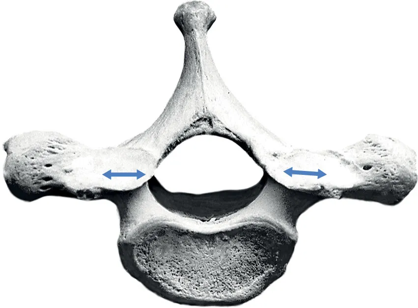
Figure 1.3
The superior aspect of the 1st thoracic vertebra. The zygapophyseal joints lie in the coronal plane.
Regions and osteology of the thorax
Vertebromanubrial region
There are two thoracic rings in the vertebromanubrial region (upper thorax) and both have a direct attachment to the manubrium. This region contains the 1st and 2nd thoracic vertebrae, the 1st and 2nd ribs, and the manubrium.
The 1st thoracic vertebra has a large, nonbifid spinous process and the superior aspect of this process is in the same transverse plane as the T1–T2 zygapophyseal joints (Fig. 1.1B). The facets on the superior articular processes, which articulate with C7, are in the coronal plane (Fig. 1.3) while those on the inferior articular process, which articulate with T2, present a slight curve in both the transverse and sagittal planes (Fig. 1.4). The transverse processes of T1 are long and thick and are located between the superior and inferior articular processes at the dorsal aspect of the pedicle. On the ventral aspect of each transverse process...