Chapter 1
Highlights of fascial anatomy, morphology and function
Robert Schleip and Werner Klingler
Fascia: more than an inert packing organ
After several decades of a Cinderella-like neglect, fascia has entered the limelight within the field of human life sciences. While literally thrown away in most anatomical dissections, this colorless fibrous tissue has mostly been treated as a dull and inert packaging organ. There are several reasons for this neglect, one of which is the lack of clear distinctions, based on the ubiquitous and seemingly disordered nature of this tissue, compared with the shiny muscles and organs underneath. Another and more important reason for the severe neglect of scientific attention concerned the lack of adequate measurement tools. While x-ray imaging allowed a detailed study of bones and the electromyography of muscles, for many decades, changes in fascia were difficult to measure. For example, the fascia lata or lumbar fascia is typically less than 2 mm thick, and a local increase in thickness of 20% was too small to be seen by ultrasound (or any other affordable imaging technology in clinical practice), although it may be easily palpable to the hand of a therapist and may also be felt by the client during movement.
This unfortunate situation has changed significantly in recent years. Advances in ultrasound measurement, as well as in histology, have contributed to an increase in fascia-related studies (Chaitow et al., 2012). Examples of some of the novel insights within the rapidly advancing field of international fascia research include the role of the human thoracolumbar fascia as a potential source of low back pain (Wilke et al., 2017), the discovery of fasciacytes as a new type of connective tissue cell (see Chapter 9), and the recognition of a close link between the sympathetic nervous system and active fascial tonicity (Schleip et al., 2019). Clinical fields whose practitioners have an avid interest and participation in this process include manual therapies, physiotherapy, scar treatment, oncology (based on the matrix-dependent behavior of cancer cells), surgery and rehabilitative medicine. Similarly, sports science is embracing these developments. The first congresses on Connective tissues in sports medicine, hosted at Ulm University in 2013 and 2017, served as an important impetus for the development of this field. Today, fascia has become a favored new subject in conferences for sports sciences as well as among movement teachers.
“A fascia” and “the fascial system”
Most classical anatomy textbooks contain several descriptions of specific fasciae with clear definitions of its anatomical position and main functions. Examples include Osborne’s fascia as an outer sheath of the cubital tunnel or the thoracolumbar fascia covering back muscles and spanning from spine to pelvis. However, only those membranous connective tissue structures that could easily be dissected with a conventional scalpel (and without use of a microscope) were labeled fasciae. Other collagenous structures, such as tendons, aponeuroses or the small tubular endomysium layers within the intramuscular connective tissue, were excluded. Due to the diverse form and function of fasciae, it is impossible to conclusively define the fascia. However, there are morphological and functional key elements which have been identified as common properties of a fascia. Since the use of the term fascia is non-uniform, an expert Fascia Nomenclature Committee, consisting of anatomists, physiologists, biologists and clinicians, was established in 2014.
According to this committee, the new functional terminology (see the box below) defines fascia as all components that form an interconnected collagenous three-dimensional continuum of soft collagen containing loose and dense fibrous connective tissues that permeate the body (Schleip et al., 2019).
Functional definition of the fascial system
The fascial system consists of the three-dimensional continuum of soft collagen containing loose and dense fibrous connective tissues that permeate the body.
It incorporates elements such as adipose tissue, adventitiae and neurovascular sheaths, aponeuroses, deep and superficial fasciae, epineurium, joint capsules, ligaments, membranes, meninges, myofascial expansions, periostea, retinacula, septa, tendons, visceral fasciae and all the intramuscular and intermuscular connective tissues, including endomysium/perimysium/epimysium.
The fascial system surrounds, interweaves between and interpenetrates all organs, muscles, bones and nerve fibers, endowing the body with a functional structure and providing an environment that enables all body systems to operate in an integrated manner.
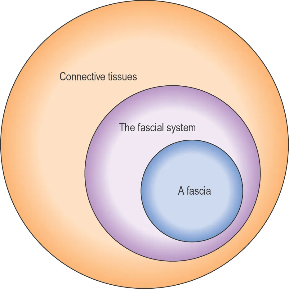
While a narrower fascia terminology is still recommended for more detail-oriented descriptions in the classical fields of histology and microscopic anatomy (see the box below), the expert panel suggests using this new and wider definition for the description and discussion of functional fascia properties, such as force transmission, sensory capacities, fibrotic pathologies or wound healing. Congruently, this will be the terminology used for most of the subsequent chapters in this book. While all connective tissues—including, for example, cartilage and bone—are derivatives of the embryological mesenchyme, only some of them are considered to be part of the fascial system, and only those with very special architecture are referred to as “proper fascia” (Figure 1.1).
Histological/anatomical definition of “a fascia”
A fascia is a sheath, a sheet or any other dissectible aggregation of connective tissue that forms beneath the skin to attach, enclose and separate muscles and other internal organs.
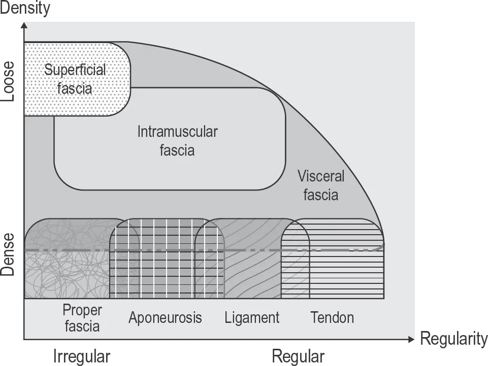
In terms of force transmission, the expression of a collagen fiber network tends to be associated with repeated tensional strain demands. The specific shape of a fascial tissue depends on the local history of these tensional forces. If the local tensional demands are mostly unidirectional and have involved high loads, then the fascial net will express these in the shape of a tendon or ligament. In other circumstances, it may express them as a two-directional or multidirectional membrane, or as a loose fibrous areolar safron (Figure 1.2). The term “fascia” is, thus, fairly synonymous with the layperson’s understanding of the term “connective tissue” (although in medical science the term “connective tissue” includes bones, cartilage and even blood, all of which derive from the embryonic mesenchyme tissue).
Figure 1.1
The nomenclature recommendations of the Fascial Nomenclature Committee are based on the understanding that the wider and more functional term, “the fascial net” (which some authors replace with “fascial tissues”), describes a subset of tissues belonging to the connective tissue system of the body. Similarly, the term, “a fascia” (also called “proper fascia” by some authors), describes a subset of tissues within the larger category of “the fascial system”.
Illustration from Schleip et al. (2019).
A body-wide interconnected tensional network
An advantage of this new and more encompassing terminology is that it recognizes the widespread continuities of this fibrous network, while still allowing for a detailed description of the local architecture. Note that, in contrast to the simplified anatomical textbook illustrations, the collagenous tissues around major joints in the human body express large areas of gradual transition, where a clear distinction between ligament, capsule, tendon, septum or muscular envelope is virtually impossible.
Figure 1.2 Different connective tissues as specializations of the global fascial net
In the new functional terminology, all collagenous fibrous connective tissues are considered as being part of the fascial system. These tissues differ in terms of their density and the directional alignment of collagen fibers. For example, superficial fascia is characterized by a relatively low density and mostly multidirectional or irregular fiber alignment, whereas in the denser tendons or ligaments, the fibers are mostly unidirectional. Note that the intramuscular fasciae—septi, perimysium and endomysium—may express varying degrees of directionality and density. The same is true for the visceral fasciae, like the very soft greater omentum in the belly or the much tougher pericardium. Depending on local loading history, fascia proper can express a unidirectional lattice-like or multidirectional arrangement.
Illustration courtesy of fascialnet.com.
Force transmission from the muscle to the skeleton also involves more extramuscular myofascial delineations than was classically assumed. This insight constitutes a challenge to the classical biomechanical concepts in musculoskeletal medicine, in which many experts considered the mechanical importance of fascial envelopes to be roughly similar to the wrapping of an average gift in relation to its content. Just as most gifts function equally well without their wrapping, it had been considered that the function of a muscle could be understood when studying its origin, insertion and myofiber orientation in a fascia-free condition. In contrast to this common assumption, the extensive work of Huijing (2007) has shown impressively how muscles transmit up to 40% of their contraction force not into their respective tendon, but rather via fascial connections into other muscles that are positioned next to them. Interestingly, this often involves force transmission to antagonistic muscles, which are then co-stiffened and tend to increase resistance to this primary movement. An increase of this particular force transmission to antagonistic muscles has been shown to be an important complication in many spastic contractures (Huijing, 2007).
Important muscular force transmissions, via their fascial connections, have been shown between:
•latissimus dorsi and contralateral gluteus maximus via the lumbodorsal fascia (Barker et al., 2004);
•biceps femoris to the erector spinae fascia via the sacrotuberous ligament (Vleeming et al., 1995);
•biceps brachii and the flexor muscles of the lower arm via the lacertus fibrosis (Brasseur, 2012);
•gluteus maximus and lower leg muscles via the fascia lata (Stecco et al., 2013).
Ingber (1998) showed that the architecture of cells can be understood to behave like a tensegrity structure. In a tensegrity structure the compressional elements (struts) are suspended without any compressional contact towards each other, whereas the tensional elements (elastic bands or membranes) are all connected with one another in a global tension-transmitting network. This model served as a basic inspiration for the field of fascia research. Driven by the observation that healthy human bodies express a higher degree of tensegrity-like qualities in their movements, many clinicians as well as scientists have started to see the fascial web as the elastic elements of a tensegrity structure, in which bones and cartilage are suspended as spacers, rather than as classical weight-bearing structures (Levin, 2003). While this is based on the assumption that the human body is a pure tensegrity structure, it has been argued that a proper understanding of the complex force transmission dynamics in the human body should also include rheological (fluid sheer motion-orientated) properties in which sponge-like motion resistance patterns play important contributing factors as well (Bordoni et al., 2019). Nevertheless, the above examples of myofascial force transmission across several joints show that a tensegrity-inspired perspective offers an improved understanding of the fascial net and its role in musculoskeletal dynamics.
Components of fascial tissues
Fascial connective tissues basically consist of two components, cells plus the extracellular matrix (Figure 1.3). Unlike most other tissues, the cells take up a very minor part of the total volume (usually less than 5%). Most of the cells are fibroblasts, which function as construction and maintenance workers for the surrounding matrix. The matrix consists of two parts: ground substance and fibers. The ground substance consists mostly of water, which is bound by proteoglycans. Most of the fibers are col...