Handbook of Oral Pathology and Oral Medicine
S. R. Prabhu
- English
- ePUB (mobile friendly)
- Available on iOS & Android
Handbook of Oral Pathology and Oral Medicine
S. R. Prabhu
About This Book
Handbook of Oral Pathology and Oral Medicine
Discover a concise overview of the most common oral diseases in a reader-friendly book
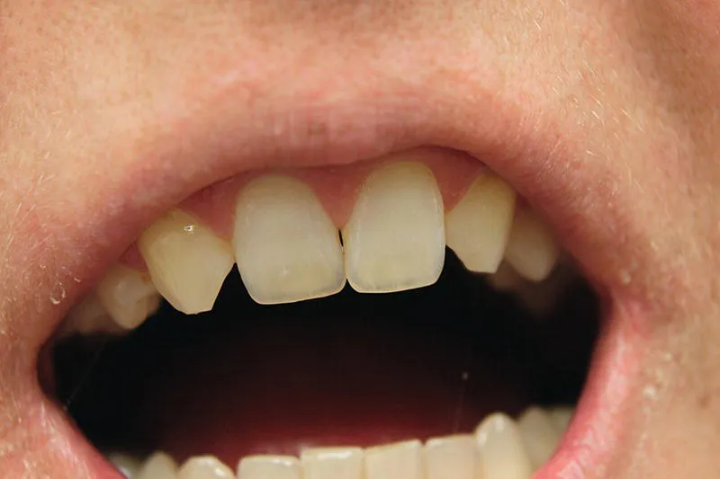
Handbook of Oral Pathology and Oral Medicine delivers a succinct overview of a range of oral diseases. The book contains up-to-date evidence-based information organized by clinical topic and supported by over 300 clinical, radiological, and microscopic images. Each chapter includes topics following universally respected curricula of oral pathology and oral medicine.
Divided into seven parts, it covers core topics such as pathology of teeth, pulp, and supporting structures, pathology of jawbones, pathology of the oral mucosa, pathology of the salivary glands, clinical presentation of mucosal disease, orofacial pain, and miscellaneous topics of clinical relevance.
Written for undergraduate dental students, dental hygienists and oral health therapists, Handbook of Oral Pathology and Oral Medicine is an ideal quick reference and is also useful to dental educators and practitioners.
Frequently asked questions
Information
Part I
Pathology of Teeth and Supporting Structures
1
Disorders of Tooth Development and Eruption
CHAPTER MENU
- 1.1 Anodontia, Hypodontia and Oligodontia
- 1.2 Hyperdontia (Supernumerary Teeth)
- 1.3 Microdontia and Macrodontia
- 1.4 Gemination, Fusion and Concrescence
- 1.5 Taurodontism and Dilaceration
- 1.6 Amelogenesis Imperfecta
- 1.7 Dentinogenesis Imperfecta
- 1.8 Dentinal Dysplasia (Dentin Dysplasia)
- 1.9 Regional Odontodysplasia (Ghost Teeth)
- 1.10 Delayed Tooth Eruption
- 1.11 Tooth Impaction (Impacted Teeth)
- 1.12 Dens Invaginatus and Dens Evaginatus
- 1.13 Fluorosis (Mottled Enamel)
- 1.14 TetracyclineâInduced Discolouration of Teeth: Key Features
- 1.15 Enamel Pearl: Key Features
- 1.16 Talon Cusp: Key Features
- 1.17 Hutchinson's Incisors and Mulberry Molars: Key Features
- 1.18 Tooth Ankylosis: Key Features
- 1.19 Supernumerary Roots: Key Features
1.1 Anodontia, Hypodontia and Oligodontia
1.1.1 Definition/Description
- Anodontia: All teeth are developmentally (congenitally) missing
- Hypodontia: one to six developmentally (congenitally) missing teeth except the third molars
- Oligodontia: more than six developmentally (congenitally) missing teeth
1.1.2 Frequency
- Anodontia: extremely rare
- Hypodontia: incidence: 4.4â13.4%. The most common developmental anomaly
- Oligodontia: incidence: 0.25%
- Hypodontia of the deciduous teeth affects less than 1% of children
1.1.3 Aetiology/Risk Factors
- Anodontia: mutations in EDA, EDAR and EDARADD genes
- Hypodontia: mutations in MSX1, PAX9, IRF6, GREM2, AXIN2, LRP6, SMOC2, LTBP3, PITX2 and WNT10B. WNT10A genes
- Oligodontia: mutations in MSX1, PAX9, IRF6, GREM2, AXIN2, LRP6, SMOC2, LTBP3, PITX2 and WNT10B. WNT10A genes
- Environmental factors: hypodontia can result from trauma to the developing dental tissues such as chemotherapy or radiotherapy
1.1.4 Clinical Features
- The most common missing multiple teeth (other than third molars) are:
- Maxillary lateral incisors (Figure 1.1). This is followed by the mandibular second premolars and maxillary second premolars
- Skin/nails/hair/sweat glands may be affected in some cases of hypodontia
- Morphology of teeth may be defective (micr...