Consumer health information about diagnosis, treatment, and management of Parkinson disease and other hypokinetic and hyperkinetic movement disorders, along with advice for family members and caregivers.
- English
- ePUB (mobile friendly)
- Available on iOS & Android
eBook - ePub
Movement Disorders Sourcebook, 4th Ed.
About this book
Trusted by 375,005 students
Access to over 1.5 million titles for a fair monthly price.
Study more efficiently using our study tools.
Information
Topic
MedicineSubtopic
General HealthPart 1 | Introduction to Movement Disorders
Chapter 1 | Anatomy of the Brain: How the Brain Controls the Body’s Movements
The central nervous system (CNS) consists of the brain and spinal cord, which are located in the dorsal body cavity. The brain is surrounded by the cranium, and the spinal cord is protected by the vertebrae. The brain is continuous with the spinal cord at the foramen magnum. In addition to bone, the CNS is surrounded by connective tissue membranes, called “meninges,” and by cerebrospinal fluid. The following are the major components of the brain and CNS:
- Neurons and glial cells
- Brain
- Meninges
- Spinal cord
- Cranial nerves
- Pineal and pituitary glands
Brain
Cerebrum
The cerebrum is the part of the brain that receives and processes conscious sensation, generates thought, and controls conscious
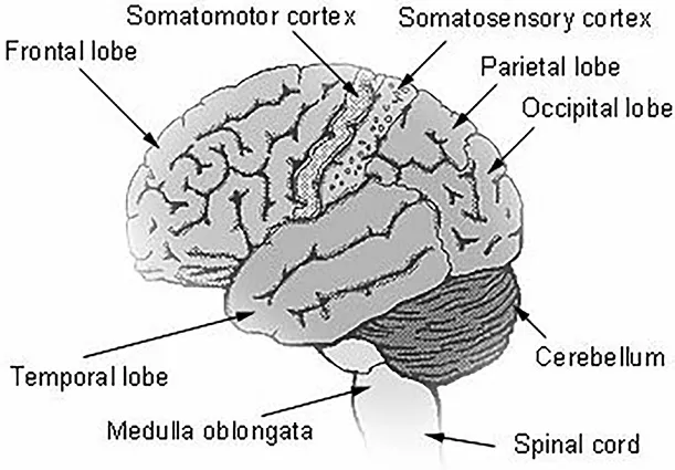
Figure 1.1. Lobes of the Cerebrum
activity. It is the uppermost and largest part of the brain and is divided into left and right hemispheres, which are joined by and communicated through the corpus callosum.
Each cerebral hemisphere is divided into five lobes, four of which have the same name as the bone over them: the frontal lobe, the parietal lobe, the occipital lobe, and the temporal lobe. A fifth lobe, the insula or island of Reil, lies deep within the lateral sulcus.
Cerebellum
The cerebellum is a cauliflower-shaped part of the brain located in the hindbrain, at the bottom rear of the head, directly behind the pons. The cerebellum is a complex system mostly dedicated to the intricate coordination of voluntary movement, including walking and balance. Damage to the cerebellum leaves the sufferer with a gait that appears drunken and is difficult to control.
Ventricles and Cerebrospinal Fluid
A series of interconnected, fluid filled cavities called “ventricles” lie within the brain. The fluid is cerebrospinal fluid (CSF), which also circulates over the outside of the brain and spinal cord.
Brainstem
The brainstem is the part of the brain continuous with the spinal cord and comprising the medulla oblongata, pons, midbrain, and parts of the hypothalamus.
Tentorium
The tentorium is a fold of the dura mater, which separates the cerebellum from the cerebrum and often encloses a process or plate of the skull called the “bony tentorium.”
Meninges
There are three layers of meninges around the brain and spinal cord. The outer layer, the dura mater, is tough, white fibrous connective tissue. The middle layer of meninges is the arachnoid, a thin layer resembling a cobweb with numerous thread-like strands attaching it to the innermost layer. The space under the arachnoid, the subarachnoid space, is filled with cerebrospinal fluid and contains blood vessels. The pia mater is the innermost layer of meninges. This thin, delicate membrane is tightly bound to the surface of the brain and spinal cord and cannot be dissected away without damaging the surface.
Spinal Cord
The spinal cord extends from the foramen magnum at the base of the skull to the level of the first lumbar vertebra. The cord is continuous with the medulla oblongata at the foramen magnum. Like the brain, the spinal cord is surrounded by bone, meninges, and cerebrospinal fluid.
The spinal cord is divided into 31 segments, with each segment giving rise to a pair of spinal nerves. At the distal end of the cord,
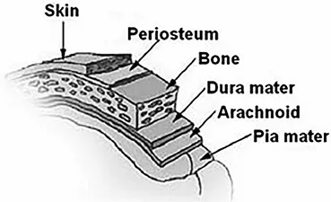
Figure 1.2. Meninges
Dura matter – outer layer lining skull
Arachnoid (matter) – contains blood vessels
Subarachnoid space – filled with CSF
Pia mater – covers brain
many spinal nerves extend beyond the conus medullaris to form a collection that resembles a horse’s tail. This is the cauda equina. In cross section, the spinal cord appears oval in shape.
_____________
This chapter includes text excerpted from “Anatomy and Function Areas of the Brain and CNS,” Surveillance, Epidemiology and End Results Program (SEER), National Cancer Institute (NCI), September 7, 2016. Reviewed July 2021.
Chapter 2 | Anatomy of the Nervous System
The nervous system is the major controlling, regulatory, and communicating system in the body. It is the center of all mental activity including thought, learning, and memory. Together with the endocrine system, the nervous system is responsible for regulating and maintaining homeostasis. Through its receptors, the nervous system keeps us in touch with our environment, both external and internal.
Like other systems in the body, the nervous system is composed of organs, principally the brain, spinal cord, nerves, and ganglia. These, in turn, consist of various tissues, including nerve, blood, and connective tissue. Together these carry out the complex activities of the nervous system.
The various activities of the nervous system can be grouped together as three general, overlapping functions:
- Sensory
- Integrative
- Motor
Millions of sensory receptors detect changes, called “stimuli,” which occur inside and outside the body. They monitor such things as temperature, light, and sound from the external environment. Inside the body, the internal environment, receptors detect variations in pressure, pH, carbon dioxide concentration, and the levels of various electrolytes. All of this gathered information is called “sensory input.”
Sensory input is converted into electrical signals called “nerve impulses” that are transmitted to the brain. There the signals are brought together to create sensations, to produce thoughts, or to add to memory; decisions are made in each moment based on the sensory input. This is integration.
Based on the sensory input and integration, the nervous system responds by sending signals to muscles, causing them to contract, or to glands, causing them to produce secretions. Muscles and glands are called “effectors” because they cause an effect in response to directions from the nervous system. This is the motor output or motor function.
Nerve Tissue
Although the nervous system is very complex, there are only two main types of cells in nerve tissue. The actual nerve cell is the neuron. It is the “conducting” cell that transmits impulses and the structural unit of the nervous system. The other type of cell is neuroglia, or glial, cell. The word “neuroglia” means “nerve glue.” These cells are nonconductive and provide a support system for the neurons. They are a special type of “connective tissue” for the nervous system.
Neurons
Neurons, or nerve cells, carry out the functions of the nervous system by conducting nerve impulses. They are highly specialized and amitotic. This means that if a neuron is destroyed, it cannot be replaced because neurons do not go through mitosis. The image below illustrates the structure of a typical neuron.
Each neuron has three basic parts: cell body (soma), one or more dendrites, and a single axon.
Dendrites
Dendrites and axons are cytoplasmic extensions, or processes, that project from the cell body. They are sometimes referred to as fibers.
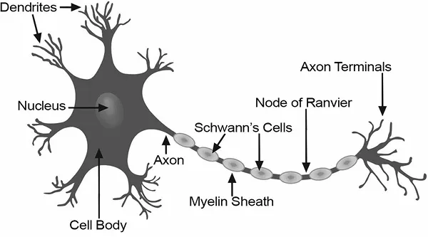
Figure 2.1. Structure of a Typical Neuron
Dendrites are usually, but not always, short and branching, which increases their surface area to receive signals from other neurons. The number of dendrites on a neuron varies. They are called “afferent processes” because they transmit impulses to the neuron cell body. There is only one axon that projects from each cell body. It is usually elongated and because it carries impulses away from the cell body, it is called an “efferent process.”
Axon
An axon may have infrequent branches called “axon collaterals.” Axons and axon collaterals terminate in many short branches or telocentric. The distal ends of the telocentric are slightly enlarged to form synaptic bulbs. Many axons are surrounded by a segmented, white, fatty substance called “myelin” or the “myelin sheath.” Myelinated fibers make up the white matter in the CNS, while cell bodies and unmyelinated fibers make the gray matter. The unmyelinated regions between the myelin segments are called the “nodes of Ranvier.”
In the peripheral nervous system, the myelin is produced by Schwann cells. The cytoplasm, nucleus, and outer cell membrane of the Schwann cell form a tight covering around the myelin and around the axon itself at the nodes of Ranvier. This covering is the neurilemma, which plays an important role in the regeneration of nerve fibers. In the CNS, oligodendrocytes produce myelin, but there is no neurilemma, which is why fibers within the CNS do not regenerate.
Functionally, neurons are classified as afferent, efferent, or interneurons (association neurons) according to the direction in which they transmit impulses relative to the central nervous system. Afferent, or sensory, neurons carry impulses from peripheral sense receptors to the CNS. They usually have long dendrites and relatively short axons. Efferent, or motor, neurons transmit impulses from the CNS to effector organs such as muscles and glands. Efferent neurons usually have short dendrites and long axons. Interneurons, or association neurons, are located entirely within the CNS in which they form the connecting link between the afferent and efferent neurons. They have short dend...
Table of contents
- Cover
- Halftitle
- Title
- Copyright
- Table of Contents
- Part 1. Introduction to Movement Disorders
- Part 2. Hypokinetic Movement Disorders
- Part 3. Hyperkinetic Movement Disorders
- Part 4. Cerebellar Disorders and Ataxias
- Part 5. Childhood Movement Disorders
- Part 6. Diagnosis and Treatment of Movement Disorders
- Part 7. Living with Movement Disorders
- Part 8. Additional Help and Information
- Index
Frequently asked questions
Yes, you can cancel anytime from the Subscription tab in your account settings on the Perlego website. Your subscription will stay active until the end of your current billing period. Learn how to cancel your subscription
No, books cannot be downloaded as external files, such as PDFs, for use outside of Perlego. However, you can download books within the Perlego app for offline reading on mobile or tablet. Learn how to download books offline
Perlego offers two plans: Essential and Complete
- Essential is ideal for learners and professionals who enjoy exploring a wide range of subjects. Access the Essential Library with 800,000+ trusted titles and best-sellers across business, personal growth, and the humanities. Includes unlimited reading time and Standard Read Aloud voice.
- Complete: Perfect for advanced learners and researchers needing full, unrestricted access. Unlock 1.5M+ books across hundreds of subjects, including academic and specialized titles. The Complete Plan also includes advanced features like Premium Read Aloud and Research Assistant.
We are an online textbook subscription service, where you can get access to an entire online library for less than the price of a single book per month. With over 1.5 million books across 990+ topics, we’ve got you covered! Learn about our mission
Look out for the read-aloud symbol on your next book to see if you can listen to it. The read-aloud tool reads text aloud for you, highlighting the text as it is being read. You can pause it, speed it up and slow it down. Learn more about Read Aloud
Yes! You can use the Perlego app on both iOS and Android devices to read anytime, anywhere — even offline. Perfect for commutes or when you’re on the go.
Please note we cannot support devices running on iOS 13 and Android 7 or earlier. Learn more about using the app
Please note we cannot support devices running on iOS 13 and Android 7 or earlier. Learn more about using the app
Yes, you can access Movement Disorders Sourcebook, 4th Ed. by James Chambers in PDF and/or ePUB format, as well as other popular books in Medicine & General Health. We have over 1.5 million books available in our catalogue for you to explore.