EMBRYOLOGY provides a concise and highly illustrated text, which confines its descriptions to those that are relevant for modern undergraduate and postgraduate medical courses, and similar courses in other related disciplines. An appreciation of embryology is essential to understand topological relationships in gross anatomy and to explain many congenital anomalies. Each chapter is supplemented by clinical point 'boxes' and by key revision points.- Text in concise Illustrated Colour Text style, so core information on embryology can be quickly recognised and digested.- Clear full colour diagrams and pictures make the embryological concepts clear and easily assimilated.- Clinical boxes highlight essential points of importance to medical students.
- 96 pages
- English
- ePUB (mobile friendly)
- Available on iOS & Android
eBook - ePub
About this book
Trusted by 375,005 students
Access to over 1.5 million titles for a fair monthly price.
Study more efficiently using our study tools.
Information
Topic
MedicinaSubtopic
Ginecologia e ostetriciaChapter 1 How does an embryo form?
The 1st week—Fertilization and formation of the blastocyst 1
The 2nd week—Implantation and formation of bilaminar embryonic disc 1
The 3rd week—Further development of the embryo and formation of trilaminar embryonic disc 3
The 4th week—Folding of the embryo 7
Human development begins when a spermatozoon fertilizes an oocyte. By definition, an embryo comprises the tissues formed once mitosis of an ovum (a fertilized oocyte) begins; thus even at the two-cell stage it is an embryo. These few cells multiply in number over an 8-week period into a fetus, by which time it will consist of many millions of cells. During the first two weeks (the pre-embryonic period) the embryo moves through the uterine tube to the uterus where it will implant. The differentiation that establishes the organ systems takes place between 3 and 8 weeks in the first 8 weeks following fertilization (the embryonic period). The period of time from the end of week 8 to full term (38 weeks) is a phase of growth and enlargement (the fetal period). The crucial phase during which there is potential for malformation is in the first 8 weeks, and this period is when the embryo is most vulnerable to environmental agents such as viruses and other teratogens. Table 1.1 summarizes the major events of the prenatal stages of development. The stages of the formation of an embryo are often described in relation to weeks of development.
Table 1.1 Stages of development before birth
| Time period | Stage | Main events |
|---|---|---|
| Conception to week 2 | Pre-embryonic period | Fertilized ovum undergoes mitosis. Formation of morula; appearance of blastocyst; blastocyst implanted. |
| 2nd to 8th week | Embryonic period | Germ layers and placenta develop. Main body systems form. |
| 9th week to birth | Fetal period | Further growth and development of organs. Locomotor system becomes functional. |

Abnormal sites of implantation can sometimes occur, due to slow transit of the ovum along the uterine tube. The most common site of ectopic implantation is the uterine tube itself, though other sites include the peritoneal cavity or on the surface of the ovary. Such embryos do not come to term because the abnormal implantation site is unable to sustain the developing embryo. Furthermore, the invasive trophoblast tissue causes haemorrhage which can be life-threatening.
The stages leading up to fertilization (including gametogenesis and the histology of the uterus at the time of implantation—see ICT Histology), however, are beyond the scope of this book.
The 1st week—Fertilization and formation of the blastocyst
A fertilized ovum has a diploid number of chromosomes and once the second meiotic division has been completed, the stage of cleavage can begin. This consists of a series of rapid mitotic cell divisions in which the ovum divides over a period of about 3 days, resulting in the so-called 16-cell-stage embryo (Figs 1.1 and 1.2A). Each cell is known as a blastomere. After each cleavage division, whilst the number of cells increases, the size of each cell diminishes. The solid sphere of cells that forms is known as a morula, because it was thought to resemble a mulberry! Each of these new daughter cells is, at this stage, pluripotential. In other words, each daughter cell has the potential to differentiate into cells of any lineage.
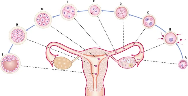
Fig. 1.1 Stages of pre-embryonic development during the first week. (A) ovulated oocyte; (B) fertilization; (C) stage of pronuclei formation; (D) first cleavage spindle; (E–G) cleavage of zygote; (H) morula; (I) blastocyst formation.
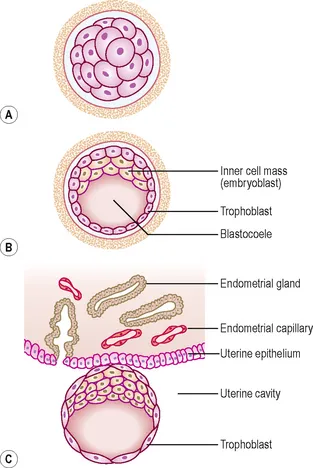
Fig. 1.2 Early stages of implantation. A 3-day morula (A) and sections of blastocyst are shown at 5 days (B) and 6 days (C) making contact with the uterine wall.
The morula soon shows signs of further differentiation. Cavities appear within the centre of the sphere of cells, forming a blastocyst, the cavity itself being the blastocoele (Fig. 1.2B, C). Once this stage has been reached the outer layer of the blastocyst soon thins to single-cell thickness to become the trophoblast, enclosing the enlarging fluid-filled blastocyst cavity. The central group of cells move to one pole of the blastocyst (the embryonic pole) to form the inner cell mass from which the whole embryo itself will form. The trophoblast contributes to the fetal component of the placenta (Fig. 1.3).
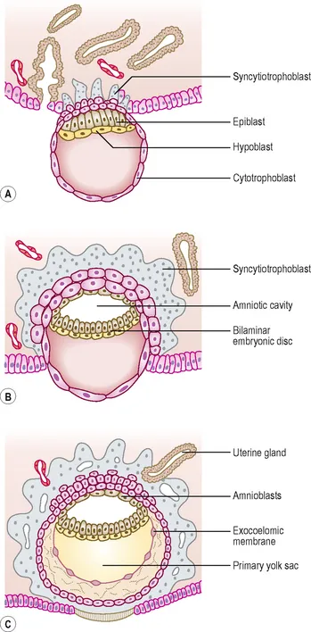
Fig. 1.3 Implantation of blastocyst. (A) A 7-day blastocyst beginning to implant. (B) By 8 days the amniotic cavity appears. (C) By 9 days the syncytiotrophoblast invades the uterine glands and capillaries.
The process of morula and blastocyst formation occurs whilst the sphere of dividing cells is in transit along the uterine tube (Fig. 1.1). Fertilization takes place in the ampulla of the uterine tube, approximately 12–24 hours after ovulation. The first mitotic division of cleavage will be completed by the time that the two-cell stage embryo reaches the middle of the tube, at about 30 hours post-fertilization. By 3 days the morula of 12–16 cells will have reached the junction of the uterine tube and the uterus. By 4–5 days the fully formed blastocyst re...
Table of contents
- Cover
- Title Page
- Front Matter
- Copyright
- Preface
- Table of Contents
- Chapter 1: How does an embryo form?
- Chapter 2: How do the placenta and fetal membranes form?
- Chapter 3: The body cavities and the diaphragm
- Chapter 4: The integumentary, skeletal and muscular systems
- Chapter 5: The respiratory system
- Chapter 6: The cardiovascular system
- Chapter 7: The digestive system
- Chapter 8: The urinary system
- Chapter 9: The reproductive system
- Chapter 10: The nervous system
- Chapter 11: Development of the head and neck, the eye and ear
- Glossary
- Subject Index
Frequently asked questions
Yes, you can cancel anytime from the Subscription tab in your account settings on the Perlego website. Your subscription will stay active until the end of your current billing period. Learn how to cancel your subscription
No, books cannot be downloaded as external files, such as PDFs, for use outside of Perlego. However, you can download books within the Perlego app for offline reading on mobile or tablet. Learn how to download books offline
Perlego offers two plans: Essential and Complete
- Essential is ideal for learners and professionals who enjoy exploring a wide range of subjects. Access the Essential Library with 800,000+ trusted titles and best-sellers across business, personal growth, and the humanities. Includes unlimited reading time and Standard Read Aloud voice.
- Complete: Perfect for advanced learners and researchers needing full, unrestricted access. Unlock 1.5M+ books across hundreds of subjects, including academic and specialized titles. The Complete Plan also includes advanced features like Premium Read Aloud and Research Assistant.
We are an online textbook subscription service, where you can get access to an entire online library for less than the price of a single book per month. With over 1.5 million books across 990+ topics, we’ve got you covered! Learn about our mission
Look out for the read-aloud symbol on your next book to see if you can listen to it. The read-aloud tool reads text aloud for you, highlighting the text as it is being read. You can pause it, speed it up and slow it down. Learn more about Read Aloud
Yes! You can use the Perlego app on both iOS and Android devices to read anytime, anywhere — even offline. Perfect for commutes or when you’re on the go.
Please note we cannot support devices running on iOS 13 and Android 7 or earlier. Learn more about using the app
Please note we cannot support devices running on iOS 13 and Android 7 or earlier. Learn more about using the app
Yes, you can access Embryology E-Book by Barry Mitchell,Ram Sharma in PDF and/or ePUB format, as well as other popular books in Medicina & Ginecologia e ostetricia. We have over 1.5 million books available in our catalogue for you to explore.