The Handbook of Small Animal Radiology and Ultrasound: Techniques and Differential Diagnoses provides a user-friendly reference for a wide range of radiographic and ultrasonographic findings in dogs and cats.Key features- Enables successful and clear interpretation of radiographs and ultrasonograms- Offers clearly sequenced text arrangement from the identification of the radiographic or sonographic abnormalities to a list of subsequent considerations for each sign- Prioritizes different clinical findings to tailor further diagnostic tests or therapeutic interventions- Takes imaging abnormalities from the descriptive to the interpretative New to this edition- Colour throughout enhances user-friendliness- Many new conditions- Extra illustrations show techniques and normal anatomy- Additional information on techniques, normal appearance and disease processes- Expanded Further Reading sections This book is intended for all users of small animal diagnostic imaging, from radiologists through to general practitioners to veterinary students, and will be an invaluable supplement to existing references in the subject.
eBook - ePub
Handbook of Small Animal Radiological Differential Diagnosis E-Book
Handbook of Small Animal Radiological Differential Diagnosis E-Book
- 382 pages
- English
- ePUB (mobile friendly)
- Available on iOS & Android
eBook - ePub
Handbook of Small Animal Radiological Differential Diagnosis E-Book
Handbook of Small Animal Radiological Differential Diagnosis E-Book
About this book
Trusted by 375,005 students
Access to over 1.5 million titles for a fair monthly price.
Study more efficiently using our study tools.
Information
Chapter 1. Skeletal system
general
CHAPTER CONTENTS
General1
1.1 Radiographic technique for the skeletal system1
1.2 Anatomy of bone: general principles2
1.3 Ossification and growth plate closures3
1.4 Response of bone to disease or injury3
1.5 Patterns of focal bone loss (osteolysis)4
1.6 Patterns of osteogenesis: periosteal reactions6
1.7 Principles of interpretation of skeletal radiographs7
1.8 Features of aggressive versus non-aggressive bone lesions8
1.9 Fractures: radiography, classification and assessment of healing9
Bones14
1.10 Altered shape of long bones14
1.11 Dwarfism15
1.12 Delayed ossification or growth plate closure15
1.13 Increased radiopacity within bone16
1.14 Periosteal reactions18
1.15 Bony masses19
1.16 Osteopenia21
1.17 Coarse trabecular pattern22
1.18 Osteolytic lesions22
1.19 Expansile osteolytic lesions24
1.20 Mixed osteolytic–osteogenic lesions25
1.21 Multifocal diseases28
1.22 Lesions affecting epiphyses29
1.23 Lesions affecting physes30
1.24 Lesions affecting metaphyses31
1.25 Lesions affecting diaphyses33
GENERAL
1.1. RADIOGRAPHIC TECHNIQUE FOR THE SKELETAL SYSTEM
The skeletal system lends itself well to radiography, but it must be remembered that only the mineralized components of bone are visible. The osteoid matrix of bone is of soft tissue radiopacity and cannot be assessed radiographically; this makes up 30–35% of adult bone. Articular cartilage is also of soft tissue opacity and is not seen on survey radiographs (see arthrography, 2.1). Lesions in the skeletal system may be radiographically subtle, and so attention to good radiographic technique is essential.
1. Highest definition film–screen combination consistent with the thickness of the area or appropriate digital radiography algorithm.
2. No grid is necessary except for the proximal limbs and spine in larger dogs; in smaller joints, insufficient scattered radiation is produced to warrant the use of a grid, and the presence of grid lines may obscure fine detail.
3. Accurate positioning and centring with a small object–film distance to minimize geometric distortion and blurring due to the penumbra effect.
4. Close collimation to enhance radiographic definition by minimizing scatter, and for radiation safety.
5. Correct exposure factors to allow examination of soft tissues as well as bone.
6. Beware of hair coat debris creating artefactual shadows.
7. Radiograph the opposite limb for comparison if necessary.
8. Use wedge filtration techniques if a whole limb view is required (e.g. for angular limb deformity); use a special wedge filter or intravenous fluid bags.
9. Optimum viewing conditions – dry films, darkened room, bright light and dimmer facility, glare around periphery of film masked off.
10. Use a magnifying glass for fine detail; use bone specimens, a film library and radiographic atlases.
11. For analogue film, ensure good processing technique to optimize contrast and definition.
12. With digital radiography, manipulation of image size and greyscale is readily performed.
1.2. ANATOMY OF BONE: GENERAL PRINCIPLES (Fig. 1.1A and B)
Apophysis – Non-articular bony protuberance for attachment of tendons and ligaments; a separate centre of ossification.
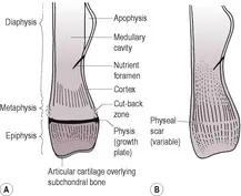 |
| Figure 1.1 (A) Anatomical features of an immature long bone; (B) anatomical features of a mature long bone. |
Articular cartilage – Soft tissue opacity, therefore appears radiolucent compared with bones (unless mineralizing through disease). Provides longitudinal growth of epiphyses.
Cancellous bone – Spongy bone consisting of a meshwork of bony trabeculae; found in epiphyses, metaphyses and small bones. A coarse trabecular pattern is seen where forces are constant and a fine trabecular pattern where they are variable. The greater surface area compared with cortical bone results in a 40 times greater rate of remodelling in response to disease or injury. The cancellous bone of skull is called diploë.
Cortex – Compact lamellar bone formed by intramembranous ossification from periosteum. Uniformly radiopaque. Thickest where the circumference of the bone is smallest, where attached soft tissues exert stress or on the concave side of a curved bone and taper to nothing in the metaphyseal region.
Diaphysis – The shaft of a long bone; a tube of cortical bone surrounding medullary cavit...
Table of contents
- Cover Image
- Table of Contents
- Front matter
- Copyright
- Foreword
- Preface
- Acknowledgements
- Chapter 1. Skeletal system
- Chapter 2. Joints
- Chapter 3. Appendicular skeleton
- Chapter 4. Head and neck
- Chapter 5. Spine
- Chapter 6. Lower respiratory tract
- Chapter 7. Cardiovascular system
- Chapter 8. Other thoracic structures
- Chapter 9. Other abdominal structures
- Chapter 10. Gastrointestinal tract
- Chapter 11. Urogenital tract
- Chapter 12. Soft tissues
- Appendix
- Index
Frequently asked questions
Yes, you can cancel anytime from the Subscription tab in your account settings on the Perlego website. Your subscription will stay active until the end of your current billing period. Learn how to cancel your subscription
No, books cannot be downloaded as external files, such as PDFs, for use outside of Perlego. However, you can download books within the Perlego app for offline reading on mobile or tablet. Learn how to download books offline
We are an online textbook subscription service, where you can get access to an entire online library for less than the price of a single book per month. With over 1.5 million books across 990+ topics, we’ve got you covered! Learn about our mission
Look out for the read-aloud symbol on your next book to see if you can listen to it. The read-aloud tool reads text aloud for you, highlighting the text as it is being read. You can pause it, speed it up and slow it down. Learn more about Read Aloud
Yes! You can use the Perlego app on both iOS and Android devices to read anytime, anywhere — even offline. Perfect for commutes or when you’re on the go.
Please note we cannot support devices running on iOS 13 and Android 7 or earlier. Learn more about using the app
Please note we cannot support devices running on iOS 13 and Android 7 or earlier. Learn more about using the app
Yes, you can access Handbook of Small Animal Radiological Differential Diagnosis E-Book by Ruth Dennis,Robert M. Kirberger,Frances Barr,Robert H. Wrigley in PDF and/or ePUB format, as well as other popular books in Medicine & Veterinary Medicine. We have over 1.5 million books available in our catalogue for you to explore.