This classic resource by Drs. Mitchel P. Goldman, Robert A Weiss, and Jean-Jerome Guex provides highly practical, up-to-date guidance for the effective management of varicose veins and other vascular anomalies. It is an indispensable reference for a wide audience including dermatologists, invasive radiologists, family practitioners, vascular and cosmetic surgeons. Clearly written by global experts, Sclerotherapy, 6th Edition, helps those new to the field to gain a firm understanding of successful techniques, as well as showing seasoned practitioners how to improve and hone their skills with today's best and newest approaches. Case studies and detailed color illustrations offer step-by-step visual guidance.- Covers everything you need to know with a practical approach, from the pathogenesis of varicosities to diagnostic and treatment options, including evidence-based decision making.- Helps you optimize outcomes and improve your surgical, injection, and laser techniques with comprehensive, visual guidance, including coverage of common pitfalls and "tricks of the trade."- Consult this title on your favorite e-reader, conduct rapid searches, and adjust font sizes for optimal readability.- Features hot topic coverage of endovenous glue and new endovenous ablation techniques, as well as updated techniques for optimal use of foam sclerotherapy and uses for solutions recently available on the market.- Discusses new concepts for treating areas other than the legs, including rejuvenation of the hands and chest.
eBook - ePub
Sclerotherapy E-Book
Treatment of Varicose and Telangiectatic Leg Veins (Expert Consult)
- 448 pages
- English
- ePUB (mobile friendly)
- Available on iOS & Android
eBook - ePub
Sclerotherapy E-Book
Treatment of Varicose and Telangiectatic Leg Veins (Expert Consult)
About this book
Trusted by 375,005 students
Access to over 1.5 million titles for a fair monthly price.
Study more efficiently using our study tools.
Information
Topic
MedicineSubtopic
Dermatology1
Anatomy
Stefano Ricci
Introduction
The anatomy chapter in a modern text devoted to sclerotherapy is traditionally not the most fascinating aspect, as the anatomy rarely changes and is very similar to that described in older texts. Anatomy chapters are rarely consulted because readers believe they know the basics of venous anatomy, but they should be reviewed regularly, and as one uses duplex ultrasound, the importance of understanding anatomy increases greatly. While this chapter reports on the images and the concepts of the classic anatomy texts that we used during our university medical studies, it is clear from our experience with duplex ultrasound observations that a ‘déjà vu’ sensation to anatomy is not entirely correct and anatomy is more than a fixed science—new understanding has been added.
Dissection anatomy, indeed, had its fullest expression from the late eighteenth to the early twentieth century (Mascagni, Gray, Sobotha, Testut, etc.) when all the aspects of dissection anatomy were definitively studied (Fig. 1.1). In the past 50 years, anatomical dissection has been little used to investigate venous anatomy, probably because of the assumption that there is nothing new to discover (but also because it is more and more difficult to find cadavers for this purpose). Meanwhile, most anatomical, clinical and surgical textbooks describe the superficial veins of the lower limb as a simple ‘tree’ formed by a few constant and recognizable veins, though clinical experience often shows anomalies and variations with respect to the classical anatomical description or even the complete absence of some of these veins. Furthermore, studies in the field of limb veins usually concern subjects with varicose pathology and rarely subjects with a normal venous system.
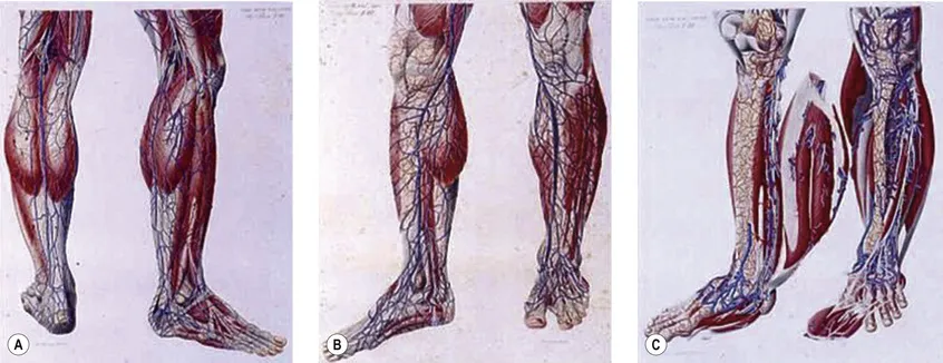
Figure 1.1 Three plates from the Piccola Anatomia, which was published in a reduced size because of the high printing costs of the time, are shown here. These demonstrate that anatomical knowledge was already complete 200 years ago. (The Grande Anatomia of Paolo Mascagni was published between 1823 and 1831 by Nicolò Capurro in Pisa).
Confirming this, the official Anatomical Terminology (Nomina Anatomica)1 includes only a limited number of veins and does not take into account their numerous variations. Inadequacy of official anatomy has caused many authors to name single veins independently or even after the author's name, which, in the absence of an accepted interpretation frame, has added some confusion. The nomenclature consensus statement of 2001 at the Rome UIP World Congress was organized with the purpose of solving this problem (see Table 1.1).2
Table 1.1
Summary of Important Changes in Nomenclature of Lower Extremity Veins
| Old Terminology | New Terminology |
| Femoral vein | Common femoral vein |
| Superficial femoral vein | Femoral vein |
| Sural veins | Sural veins Soleal veins Gastrocnemius veins (medial and lateral) |
| Hunterian perforator | Mid-thigh perforator |
| Cockett perforators | Paratibial perforator Posterior tibial perforators |
| May perforator | Ankle lateral and medial perforators |
| Gastrocnemius point | Intergemellar perforator |
Contrast phlebography, until recently the ‘gold standard’ for venous investigation, has the major drawback of being practically never complete, but rather showing only the veins filled by contrast media. Furthermore, it focuses mainly on deep veins and in pathologic conditions, and thus has not contributed much to the understanding of normal vein anatomy.
Understanding of vein anatomy did not progress much until ultrasound imaging (USI), specifically duplex scanning (DS), became an established technique for clinical investigation of patients with venous diseases. Technology simplifications and low costs have allowed its widespread use.
Ultrasound imaging makes it easy to observe the veins of the lower limb, unlike anatomical dissection and phlebography. Examination is noninvasive, repeatable and relatively low in cost. Veins can be observed at full distention, with the patient in a standing position, so that, unlike with anatomical dissection, their real volumetric relationship with the surrounding tissue is readily appreciated. Ultrasound images show not only the veins (as contrast phlebography does), but their relation to surrounding anatomical structures, in particular muscle and fascial layers. This allows precise anatomical identification of the observed veins (Fig. 1.2). Therefore, USI is a unique tool for the study of vein anatomy [ultrasound (US) dissection] and makes it possible to verify data obtained from anatomical dissections. In addition, DS allows the detection of blood flow in the observed veins with assessment of their function and involvement in venous pathology. Interestingly, USI was first employed for the clinical identification of pathologically changed veins. Later it was used for collecting data on normal vein anatomy.3
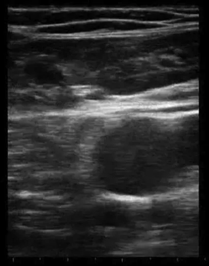
Figure 1.2 Ultrasound imaging shows the veins and their relationship to the surrounding anatomical structures, in particular other vessels, lymph nodes, bones, muscles and fascial layers. This allows precise anatomical identification of the observed veins.
In this chapter vein anatomy is first described from the traditional point of view, and successively as observed by USI with special reference to the superficial veins of the lower limb in relation to varicose vein disease and sclerotherapy. For this purpose an interpretation key is emphasized, which makes it possible to categorize the extreme variability of the superficial veins of the lower limb into a limited number of specific anatomical and varicose patterns.
Nomenclature
Nomenclature used throughout the textbook conforms to that developed at the Venous Consensus Conference Classification in 1994.4 In addition, the newest revisions of nomenclature and definitions are used, which were developed at the Nomenclature Congress in Rome in 2001 (Table 1.1).2,5 The long saphenous vein is referred to by the English-Latin term great (GSV). The short saphenous vein is referred to using the English-Latin translation small (SSV), avoiding the term ‘lesser’ as the L could be confused with the term ‘long’. Veins that ‘perforate’ the fascia are termed perforator veins. Veins that connect to other veins within a fascial plane are referred to as communicating veins. The principal deep vein of the thigh is termed the superficial femoral vein, now properly called the femoral vein. The superficial femoral vein actually has turned out to be a potentially lethal misnomer. It has been found that the use of this term is hazardous to pat...
Table of contents
- Cover image
- Title Page
- Table of Contents
- Copyright
- Video Contents
- Preface
- Additional Contributors
- Introduction
- 1 Anatomy
- 2 Adverse Sequelae and Complications of Venous Hypertension
- 3 Pathophysiology of Varicose Veins
- 4 Pathophysiology of Telangiectasias
- 5 Noninvasive Examination of the Patient Before Sclerotherapy
- 6 Use of Compression Therapy
- 7 Mechanism of Action of Sclerotherapy
- 8 Complications and Adverse Sequelae of Sclerotherapy
- 9 Clinical Methods for Sclerotherapy of Varicose Veins
- 10 Role of Surgery in the Treatment of Varicose Veins
- 11 Intravascular Approaches to the Treatment of Varicose Veins
- 12 Clinical Methods for Sclerotherapy of Telangiectasias
- 13 Treatment of Leg Telangiectasias with Laser and High-Intensity Pulsed Light
- 14 Venoactive Drugs
- Index
Frequently asked questions
Yes, you can cancel anytime from the Subscription tab in your account settings on the Perlego website. Your subscription will stay active until the end of your current billing period. Learn how to cancel your subscription
No, books cannot be downloaded as external files, such as PDFs, for use outside of Perlego. However, you can download books within the Perlego app for offline reading on mobile or tablet. Learn how to download books offline
Perlego offers two plans: Essential and Complete
- Essential is ideal for learners and professionals who enjoy exploring a wide range of subjects. Access the Essential Library with 800,000+ trusted titles and best-sellers across business, personal growth, and the humanities. Includes unlimited reading time and Standard Read Aloud voice.
- Complete: Perfect for advanced learners and researchers needing full, unrestricted access. Unlock 1.5M+ books across hundreds of subjects, including academic and specialized titles. The Complete Plan also includes advanced features like Premium Read Aloud and Research Assistant.
We are an online textbook subscription service, where you can get access to an entire online library for less than the price of a single book per month. With over 1.5 million books across 990+ topics, we’ve got you covered! Learn about our mission
Look out for the read-aloud symbol on your next book to see if you can listen to it. The read-aloud tool reads text aloud for you, highlighting the text as it is being read. You can pause it, speed it up and slow it down. Learn more about Read Aloud
Yes! You can use the Perlego app on both iOS and Android devices to read anytime, anywhere — even offline. Perfect for commutes or when you’re on the go.
Please note we cannot support devices running on iOS 13 and Android 7 or earlier. Learn more about using the app
Please note we cannot support devices running on iOS 13 and Android 7 or earlier. Learn more about using the app
Yes, you can access Sclerotherapy E-Book by Mitchel P. Goldman,Robert A Weiss in PDF and/or ePUB format, as well as other popular books in Medicine & Dermatology. We have over 1.5 million books available in our catalogue for you to explore.