Ideal for students and as a review for practicing clinicians, Goldberger's Clinical Electrocardiography explains the fundamentals of ECG interpretation and analysis, helping facilitate an understanding of rhythm disorders and the relevant clinical outcomes. The authors take readers through the nuts and bolts of ECG, using Dr. Ary Goldberger's award-winning teaching style to clarify complex concepts in an easy-to-read manner. You'll learn simple waveform analysis and beyond to present ECGs as they are used in hospital wards, outpatient clinics, emergency departments, and most especially intensive care units — where the recognition of normal and abnormal patterns is the starting point in patient care.- Includes Clinical Pearls and Review Points in each chapter, as well as indispensable self-tests on interpreting and using ECGs to formulate diagnoses.- Covers the nuts and bolts of ECG, explaining how to read the data and then interpret the subsequent clinical findings.- Features practical, comprehensive coverage of the true-to-life clinical appearance of ECGs.- Provides ECG differential diagnoses so you can answer the question, "What else could it be?"- Expert Consult eBook version included with purchase. This enhanced eBook experience allows you to search all of the text, figures, images, and references from the book on a variety of devices.- Includes new self-assessment ECGs and questions in the Expert Consult eBook.- Enhances your understanding of difficult concepts through several new illustrations and animations.- Highlights the latest information on intraventricular and atrioventricular (AV) conduction disturbances; sudden cardiac arrest; myocardial ischemia and infarction; drug toxicities; and electronic pacemakers and ICDs.
eBook - ePub
Clinical Electrocardiography: A Simplified Approach E-Book
Clinical Electrocardiography: A Simplified Approach E-Book
- 304 pages
- English
- ePUB (mobile friendly)
- Available on iOS & Android
eBook - ePub
Clinical Electrocardiography: A Simplified Approach E-Book
Clinical Electrocardiography: A Simplified Approach E-Book
About this book
Trusted by 375,005 students
Access to over 1.5 million titles for a fair monthly price.
Study more efficiently using our study tools.
Information
Topic
MedicineSubtopic
CardiologyPart I
Basic Principles and Patterns
Chapter 1
Essential Concepts
What Is an ECG?
The electrocardiogram (ECG or EKG) is a special type of graph that represents cardiac electrical activity from one instant to the next. Specifically, the ECG provides a time-voltage chart of the heartbeat. The ECG is a key component of clinical diagnosis and management of inpatients and outpatients because it may provide critical information. Therefore, a major focus of this book is on recognizing and understanding the “signature” ECG findings in life-threatening conditions such as acute myocardial ischemia and infarction, severe hyperkalemia or hypokalemia, hypothermia, certain types of drug toxicity that may induce cardiac arrest, pericardial (cardiac) tamponade, among many others.
The general study of ECGs, including its clinical applications, technologic aspects, and basic science underpinnings, comprises the field of electrocardiography. The device used to obtain and display the conventional (12-lead) ECG is called the electrocardiograph, or more informally, the ECG machine. It records cardiac electrical currents (voltages or potentials) by means of sensors, called electrodes, selectively positioned on the surface of the body.a Students and clinicians are often understandably confused by the basic terminology that labels the graphical recording as the electrocardiogram and the recording device as the electrocardiograph! We will point out other potentially confusing ECG semantics as we go along.
Contemporary ECGs are usually recorded with disposable paste-on (adhesive) silver–silver chloride electrodes. For the standard ECG recording, electrodes are placed on the lower arms, lower legs, and across the chest wall (precordium). In settings such as emergency departments, cardiac and intensive care units (CCUs and ICUs), and ambulatory (e.g., Holter) monitoring, only one or two “rhythm strip” leads may be recorded, usually by means of a few chest and abdominal electrodes.
ABCs of Cardiac Electrophysiology
Before the basic ECG patterns are discussed, we review a few simple-to-grasp but fundamental principles of the heart's electrical properties.
The central function of the heart is to contract rhythmically and pump blood to the lungs (pulmonary circulation) for oxygenation and then to pump this oxygen-enriched blood into the general (systemic) circulation. Furthermore, the amount of blood pumped has to be matched to meet the body's varying metabolic needs. The heart muscle and other tissues require more oxygen and nutrients when we are active compared to when we rest. An important part of these auto-regulatory adjustments is accomplished by changes in heart rate, which, as described below, are primarily under the control of the autonomic (involuntary) nervous system.
The signal for cardiac contraction is the spread of synchronized electrical currents through the heart muscle. These currents are produced both by pacemaker cells and specialized conduction tissue within the heart and by the working heart muscle itself.
Pacemaker cells are like tiny clocks (technically called oscillators) that automatically generate electrical stimuli in a repetitive fashion. The other heart cells, both specialized conduction tissue and working heart muscle, function like cables that transmit these electrical signals.b
Electrical Signaling in the Heart
In simplest terms, therefore, the heart can be thought of as an electrically timed pump. The electrical “wiring” of this remarkable organ is outlined in Fig. 1.1.
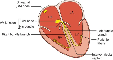
Fig. 1.1 Normally, the cardiac stimulus (electrical signal) is generated in an automatic way by pacemaker cells in the sinoatrial (SA) node, located in the high right atrium (RA). The stimulus then spreads through the RA and left atrium (LA). Next, it traverses the atrioventricular (AV) node and the bundle of His, which comprise the AV junction. The stimulus then sweeps into the left and right ventricles (LV and RV) by way of the left and right bundle branches, which are continuations of the bundle of His. The cardiac stimulus spreads rapidly and simultaneously to the left and right ventricular muscle cells through the Purkinje fibers. Electrical activation of the atria and ventricles, respectively, leads to sequential contraction of these chambers (electromechanical coupling).
Normally, the signal for heartbeat initiation starts in the pacemaker cells of the sinus or sinoatrial (SA) node. This node is located in the right atrium near the opening of the superior vena cava. The SA node is a small, oval collection (about 2 × 1 cm) of specialized cells capable of automatically generating an electrical stimulus (spark-like signal) and functions as the normal pacemaker of the heart. From the sinus node, this stimulus spreads first through the right atrium and then into the left atrium.
Electrical stimulation of the right and left atria signals the atria to contract and pump blood simultaneously through the tricuspid and mitral valves into the right and left ventricles, respectively. The electrical stimulus then spreads through the atria and part of this activation wave reaches specialized conduction tissues in the atrioventricular (AV) junction.c
The AV junction, which acts as an electrical “relay” connecting the atria and ventricles, is located near the lower part of the interatrial septum and extends into the interventricular septum (see Fig. 1.1).d
The upper (proximal) part of the AV junction is the AV node. (In some texts, the terms AV node and AV junction are used synonymously.)
The lower (distal) part of the AV junction is called the bundle of His. The bundle of His then divides into two main branches: the right bundle branch, which distributes the stimulus to the right ventricle, and the left bundle branch,e which distributes the stimulus to the left ventricle (see Fig. 1.1).
The electrical signal spreads rapidly and simultaneously down the left and right bundle branches into the ventricular myocardium (ventricular muscle) by way of specialized conducting cells called Purkinje fibers located in the subendocardial layer (roughly the inside half or rim) of the ventricles. From the final branches of the Purkinje fibers, the electrical signal spreads through myocardial muscle toward the epicardium (outer rim).
The bundle of His, its branches, and their subdivisions collectively constitute the His–Purkinje system. Normally, the AV node and His–Purkinje system provide the only electrical connection between the atria and the ventricles, unless an abnormal structure called a bypass tract is present. This abnormality and its consequences are described in Chapter 18 on Wolff–Parkinson–White preexcita...
Table of contents
- Cover image
- Title Page
- Table of Contents
- Copyright
- Dedication
- Video Contents
- Introductory Remarks
- Part I Basic Principles and Patterns
- Part II Cardiac Rhythm Disturbances
- Part III Special Topics and Reviews
- Section 1 Mini-Review Demon
- Section 2 Supplemental Extras
- Section 3 Quiz Master
- Select Bibliography
- Index
Frequently asked questions
Yes, you can cancel anytime from the Subscription tab in your account settings on the Perlego website. Your subscription will stay active until the end of your current billing period. Learn how to cancel your subscription
No, books cannot be downloaded as external files, such as PDFs, for use outside of Perlego. However, you can download books within the Perlego app for offline reading on mobile or tablet. Learn how to download books offline
Perlego offers two plans: Essential and Complete
- Essential is ideal for learners and professionals who enjoy exploring a wide range of subjects. Access the Essential Library with 800,000+ trusted titles and best-sellers across business, personal growth, and the humanities. Includes unlimited reading time and Standard Read Aloud voice.
- Complete: Perfect for advanced learners and researchers needing full, unrestricted access. Unlock 1.5M+ books across hundreds of subjects, including academic and specialized titles. The Complete Plan also includes advanced features like Premium Read Aloud and Research Assistant.
We are an online textbook subscription service, where you can get access to an entire online library for less than the price of a single book per month. With over 1.5 million books across 990+ topics, we’ve got you covered! Learn about our mission
Look out for the read-aloud symbol on your next book to see if you can listen to it. The read-aloud tool reads text aloud for you, highlighting the text as it is being read. You can pause it, speed it up and slow it down. Learn more about Read Aloud
Yes! You can use the Perlego app on both iOS and Android devices to read anytime, anywhere — even offline. Perfect for commutes or when you’re on the go.
Please note we cannot support devices running on iOS 13 and Android 7 or earlier. Learn more about using the app
Please note we cannot support devices running on iOS 13 and Android 7 or earlier. Learn more about using the app
Yes, you can access Clinical Electrocardiography: A Simplified Approach E-Book by Ary L. Goldberger,Zachary D. Goldberger,Alexei Shvilkin,Zachary D Goldberger in PDF and/or ePUB format, as well as other popular books in Medicine & Cardiology. We have over 1.5 million books available in our catalogue for you to explore.