Constituting a large percentage of everyday diagnostic practice, melanocytic pathology is a complex and challenging area with many difficult-to-diagnose lesions. This highly illustrated reference, written by three of the world's leading dermatopathologists, provides authoritative guidance in the accurate diagnosis of even the most challenging pigmented skin tumors, helping you avoid pitfalls and recognize mimics.- Covers nearly every variant of melanocytic tumors you're likely to see.- Emphasizes how to arrive at an efficient, accurate diagnosis, and includes dermoscopic findings for optimal diagnostic precision.- Discusses modern analytic techniques (cytogenetics, molecular studies) and how to use them for diagnosis.- Includes numerous case examples to illustrate the differential diagnoses and work-up; how to use ancillary techniques, along with their pros, cons, and limitations; and clinical follow-up.- Presents the knowledge and experience of Klaus Busam, Pedram Gerami, and Richard Scolyer, – three dermatopathologists who are globally renowned for their expertise in melanoma pathology and analysis of melanocytic tumors by modern ancillary diagnostic techniques.
- 608 pages
- English
- ePUB (mobile friendly)
- Available on iOS & Android
eBook - ePub
Pathology of Melanocytic Tumors E-Book
About this book
Trusted by 375,005 students
Access to over 1.5 million titles for a fair monthly price.
Study more efficiently using our study tools.
Information
Topic
MedicineSubtopic
DermatologySection I
Benign Cutaneous Melanocytic Proliferations
Introduction
- 1. Melanotic Macules, 2
- Klaus J. Busam
- 2. Acquired Melanocytic Nevi, 8
- Pedram Gerami and Klaus J. Busam
- 3. Congenital Melanocytic Nevi, 26
- Pedram Gerami
- 4. Spitz Nevi, 37
- Pedram Gerami and Klaus J. Busam
- 5. Blue Nevi and Dermal Melanocytosis, 61
- Klaus J. Busam
- 6. Deep Penetrating Nevi, 80
- Klaus J. Busam, Iwei Yeh, and Richard A. Scolyer
- 7. Melanocytic Nevi of Special Sites, 90
- Timothy VandenBoom and Pedram Gerami
- 8. Persistant/Recurrent Melanocytic Nevi, Traumatized Nevi, and Nevi Changing Under Therapy, 101
- Maija Kiuru, Pedram Gerami, and Klaus J. Busam
- 9. Combined Melanocytic Nevi, 112
- Klaus J. Busam and Richard A. Scolyer
- 10. Pigmented Epithelioid Melanocytoma, 124
- Artur Zembowicz, Jarish N. Cohen, and Philip E. LeBoit
1
Melanotic Macules
Klaus J. Busam
The term melanotic macule is used to refer to benign flat pigmented lesions, which are histopathologically characterized by melanin pigment deposited in basilar keratinocytes without or at times with a slight increase in the density of solitary units of junctional melanocytes. Cutaneous and mucosal melanotic macules are often labeled lentigo. The conjunctival equivalent has historically been termed acquired melanosis. In contrast to melanocytic nevi, there are no junctional nests in melanotic macules, lentigines, or benign acquired melanosis. The lesions differ from melanoma in situ clinically by a tendency toward small size and sharp circumscription and histopathologically by a low density of melanocytes. Melanotic macules are a common clinical presentation. They are benign and do not need to be treated. However, they may be biopsied when they display irregular features, are new, or are changing, to exclude melanoma in situ.
Solar Lentigo
Clinical Features
Solar lentigo manifests as a discrete pigmented macule or patch on sun-exposed skin of middle-aged or elderly individuals (Box 1.1).1 Its color may range from light tan to dark brown. A lesion's color may be uniform or heterogeneous. The size of a solar lentigo may be small (2 mm) or large (>1 cm). Its peripheral borders may be regular or ill-defined (Figs. 1.1 and 1.2). Multiple lesions of solar lentigo may coexist in the same anatomic region. They may collide with each other or with seborrheic or actinic keratoses, become confluent, and appear clinically complex. Clinical atypical lesions, which stand out from the background, prompt concerns for lentigo maligna (melanoma in situ) and are usually biopsied to assess for or exclude melanoma. Solar lentigines become more frequent and/or larger in area with age. Lesions of solar lentigo can also be found in young patients with sun-damaged skin and are common in children with xeroderma pigmentosum.
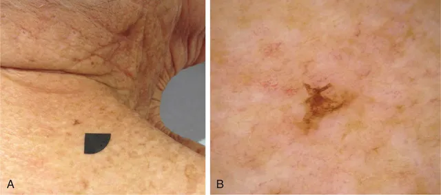
Fig. 1.1 Clinical Appearance of Solar Lentigo. (A) Pigmented macule. (B) This lesion is small but irregular in outline. Its asymmetric silhouette was the reason why it was biopsied.
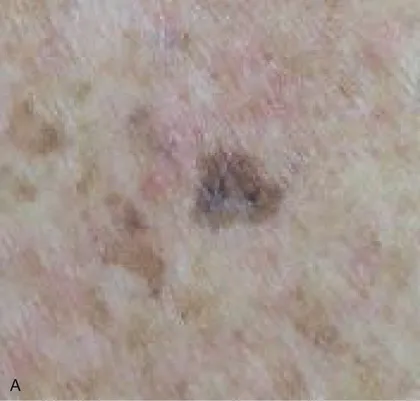
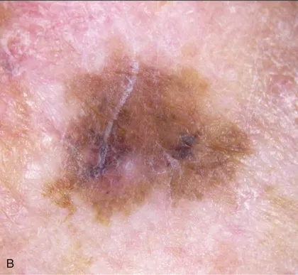
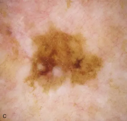
Fig. 1.2 Clinical Appearance of Solar Lentigo. (A) Hyperpigmented macule in a background of light brown macules on the chest of an elderly woman. (B) The lesion displays some irregular borders and variation in pigment intensity. (C) Corresponding dermoscopic findings.
A variant of solar lentigo clinically characterized by presentation as small dark macules, especially on the upper back, has been referred to as “ink spot” lentigo or reticulated melanotic macule.2
Histopathologic Findings
Solar lentigo is histopathologically characterized by hypermelanization of basilar keratinocytes (Figs. 1.3–1.5). Often there is associated slight epidermal hyperplasia resulting in elongated, club-shaped rete ridges. As rete ridges become longer, they may form a reticulate pattern. Lesions of solar lentigo may be associated with or develop into a seborrheic keratosis (Fig. 1.6). Pigmentation of keratinocytes is then typically most pronounced at the tip of rete ridges. In some lesions the rete ridges may...
Table of contents
- Cover image
- Title Page
- Table of Contents
- Copyright
- Dedication
- Contributors
- Preface
- Section I Benign Cutaneous Melanocytic Proliferations
- Section II Primary Cutaneous Melanoma
- Section III Primary Extracutaneous Melanocytic Proliferations
- Section IV Metastatic Melanoma
- Section V Ancillary Studies
- Section VI Prognosis, Staging, and Reporting of Melanoma
- Section VII Margin Assessment of Melanomas
- Index
Frequently asked questions
Yes, you can cancel anytime from the Subscription tab in your account settings on the Perlego website. Your subscription will stay active until the end of your current billing period. Learn how to cancel your subscription
No, books cannot be downloaded as external files, such as PDFs, for use outside of Perlego. However, you can download books within the Perlego app for offline reading on mobile or tablet. Learn how to download books offline
Perlego offers two plans: Essential and Complete
- Essential is ideal for learners and professionals who enjoy exploring a wide range of subjects. Access the Essential Library with 800,000+ trusted titles and best-sellers across business, personal growth, and the humanities. Includes unlimited reading time and Standard Read Aloud voice.
- Complete: Perfect for advanced learners and researchers needing full, unrestricted access. Unlock 1.5M+ books across hundreds of subjects, including academic and specialized titles. The Complete Plan also includes advanced features like Premium Read Aloud and Research Assistant.
We are an online textbook subscription service, where you can get access to an entire online library for less than the price of a single book per month. With over 1.5 million books across 990+ topics, we’ve got you covered! Learn about our mission
Look out for the read-aloud symbol on your next book to see if you can listen to it. The read-aloud tool reads text aloud for you, highlighting the text as it is being read. You can pause it, speed it up and slow it down. Learn more about Read Aloud
Yes! You can use the Perlego app on both iOS and Android devices to read anytime, anywhere — even offline. Perfect for commutes or when you’re on the go.
Please note we cannot support devices running on iOS 13 and Android 7 or earlier. Learn more about using the app
Please note we cannot support devices running on iOS 13 and Android 7 or earlier. Learn more about using the app
Yes, you can access Pathology of Melanocytic Tumors E-Book by Klaus J. Busam,Pedram Gerami,Richard A Scolyer in PDF and/or ePUB format, as well as other popular books in Medicine & Dermatology. We have over 1.5 million books available in our catalogue for you to explore.