Step-by-step images, board-style review questions, and coverage of new blocks make this highly respected title a must-have reference for clinical practice. Written by Andrew T. Gray, MD, PhD, one of the pioneers of the use of ultrasound to guide needle placement, Atlas of Ultrasound-Guided Regional Anesthesia, 3rd Edition, shows you how to safely and effectively use the latest methods and applications of this technique.- Helps ensure correct needle placement with numerous 3-D and long-axis views that clearly depict surrounding structures.- Includes coverage of 11 new blocks: Adductor Canal, Posterior Femoral Cutaneous, Pectoral, Quadratus Lumborum, Pudendal, Paravertebral, Transversus thoracis, Supraorbital, Transtracheal, Greater Occipital and Lesser Occipital.- Features access to 20 author-narrated videos showing proper placement of needles using ultrasound guidance, including 11 new videos: Forearm (ulnar, median and radial), Ankle (tibial, saphenous, superficial peroneal, deep peroneal, sural), Paravertebral, Adductor Canal, and Catheters.- Presents several new chapters, including Regional Anesthesia in Resource-Constrained Environments and Safety of Ultrasound Guided Regional Blocks.- Expert Consult™ eBook version included with purchase. This enhanced eBook experience allows you to search all of the text, figures, and references from the book on a variety of devices.
Trusted by 375,005 students
Access to over 1.5 million titles for a fair monthly price.
Ultrasound waves are high-frequency sound waves generated in specific frequency ranges and sent through tissues.1 How sound waves penetrate a tissue depends on the range of the frequency produced. Lower frequencies penetrate deeper than high frequencies do. The frequencies for clinical imaging (1 to 70 MHz) are well above the upper limit of normal human hearing (15 to 20 KHz). Wave motion transports energy and momentum from one point in space to another without transport of matter. In mechanical waves (e.g., water waves, waves on a string, and sound waves), energy and momentum are transported by means of disturbance in the medium because the medium has elastic properties. Any wave in which the disturbance is parallel to the direction of propagation is referred to as a longitudinal wave. Sound waves are longitudinal waves of compression and rarefaction of a medium such as air or soft tissue. Compression refers to high-pressure zones, and rarefaction refers to low-pressure zones (these zones alternate in position).
As the sound passes through tissues, it is absorbed, reflected, or allowed to pass through, depending on the echodensity of the tissue. Substances with high water content (e.g., blood, cerebrospinal fluid) conduct sound very well and reflect very poorly and thus are termed echolucent. Because they reflect very little of the sound, they appear as dark areas (hypoechoic). Substances low in water content or high in materials that are poor sound conductors (e.g., air, bone) reflect almost all the sound and appear very bright (hyperechoic). Substances with sound conduction properties between these extremes appear darker to lighter, depending on the amount of wave energy they reflect.
Audible sounds spread out in all directions, whereas ultrasound beams are well collimated. The frequency of sound does not change with propagation unless the wave strikes a moving object, in which case the changes are small. The product of the frequency and wavelength of sound waves is the wave speed. Because the speed of sound in soft tissue is nearly constant, higher-frequency sound waves have shorter wavelengths. Two adjacent structures cannot be identified as separate entities on an ultrasound scan if they are less than one wavelength apart. Therefore sound wave frequency is one of the main determinants of spatial resolution of ultrasound scans.
Chapter 2
Speed of Sound
The speed of sound is determined by properties of the medium in which it propagates. The sound velocity equals
, where B equals the bulk modulus and rho equals density. The bulk modulus is proportional to stiffness. Thus stiffness (change in shape) and wave speed are related. Density (weight per unit volume) and wave speed are inversely related. The speed of sound in a given medium is essentially independent of frequency.
Because the velocity of sound in soft tissue is 1540 m/s, 13 microseconds elapse for each centimeter of tissue the sound wave must travel (the back-and-forth time of flight). Speed-of-sound artifacts relate to both time-of-flight considerations and refraction that occurs at the interface of tissues with different speeds of sound.1-3
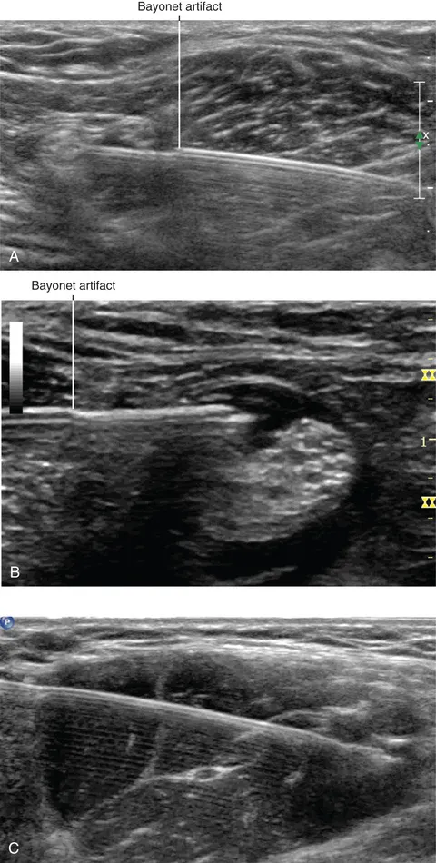
FIGURE 2.1Bayonet artifacts during popliteal block (A and B). Because the speed of sound is not necessarily homogeneous in soft tissue, the needle can sometimes appear to bend, similar to a bayonet. Actual mechanical bending of the needle typically appears as gentle bowing of the needle (C).
Chapter 3
Attenuation
Attenuation is a decrease in wave amplitude as it travels through a medium. The attenuation of ultrasound in soft tissue is approximately 0.5 to 0.75 dB/(MHz-cm), indicating that the extent of attenuation depends on the distance traveled and the frequency of insonation. The units of the attenuation coefficient directly show the greater attenuation of high-frequency ultrasound beams. In soft tissue, 80% or more of the total attenuation is caused by absorption of the ultrasound wave, thereby generating heat.
Time gain compensation (TGC) adjusts for attenuation of an ultrasound beam as a function of depth. When TGC is properly adjusted, images of similar reflectors appear the same regardless of depth.
An acoustic shadow is said to exist when a localized objec...
Table of contents
Cover image
Title Page
Table of Contents
Copyright
Dedication
Preface
Contributors
Video Contents
Section 1 Introduction to Ultrasound Imaging
Section 2 Structures
Section 3 Upper Extremity Blocks
Section 4 Lower Extremity Blocks
Section 5 Trunk Blocks
Section 6 Head and Neck Blocks
Section 7 Safety Issues
Appendix 1
Appendix 2
Appendix 3
Appendix 4
Index
Frequently asked questions
Yes, you can cancel anytime from the Subscription tab in your account settings on the Perlego website. Your subscription will stay active until the end of your current billing period. Learn how to cancel your subscription
No, books cannot be downloaded as external files, such as PDFs, for use outside of Perlego. However, you can download books within the Perlego app for offline reading on mobile or tablet. Learn how to download books offline
Perlego offers two plans: Essential and Complete
Essential is ideal for learners and professionals who enjoy exploring a wide range of subjects. Access the Essential Library with 800,000+ trusted titles and best-sellers across business, personal growth, and the humanities. Includes unlimited reading time and Standard Read Aloud voice.
Complete: Perfect for advanced learners and researchers needing full, unrestricted access. Unlock 1.5M+ books across hundreds of subjects, including academic and specialized titles. The Complete Plan also includes advanced features like Premium Read Aloud and Research Assistant.
Both plans are available with monthly, semester, or annual billing cycles.
We are an online textbook subscription service, where you can get access to an entire online library for less than the price of a single book per month. With over 1.5 million books across 990+ topics, we’ve got you covered! Learn about our mission
Look out for the read-aloud symbol on your next book to see if you can listen to it. The read-aloud tool reads text aloud for you, highlighting the text as it is being read. You can pause it, speed it up and slow it down. Learn more about Read Aloud
Yes! You can use the Perlego app on both iOS and Android devices to read anytime, anywhere — even offline. Perfect for commutes or when you’re on the go. Please note we cannot support devices running on iOS 13 and Android 7 or earlier. Learn more about using the app
Yes, you can access Atlas of Ultrasound-Guided Regional Anesthesia E-Book by Andrew T. Gray in PDF and/or ePUB format, as well as other popular books in Medicine & Anesthesiology & Pain Management. We have over 1.5 million books available in our catalogue for you to explore.