- Provides state-of-the-art coverage of CMR technologies and guidelines, including basic principles, imaging techniques, ischemic heart disease, right ventricular and congenital heart disease, vascular and pericardium conditions, and functional cardiovascular disease.- Includes new chapters on non-cardiac pathology, pacemaker safety, economics of CMR, and guidelines as well as new coverage of myocarditis and its diagnosis and assessment of prognosis by cardiovascular magnetic resonance, and the use of PET/CMR imaging of the heart, especially in sarcoidosis.- Features more than 1, 100 high-quality images representing today's CMR imaging.- Covers T1, T2 and ECV mapping, as well as T2* imaging in iron overload, which has been shown to save lives in patients with thalassaemia major- Discusses the cost-effectiveness of CMR.- Provides state-of-the-art coverage of CMR technologies and guidelines, including basic principles, imaging techniques, ischemic heart disease, right ventricular and congenital heart disease, vascular and pericardium conditions, and functional cardiovascular disease.- Includes new chapters on non-cardiac pathology, pacemaker safety, economics of CMR, and guidelines as well as new coverage of myocarditis and its diagnosis and assessment of prognosis by cardiovascular magnetic resonance, and the use of PET/CMR imaging of the heart, especially in sarcoidosis.- Features more than 1, 100 high-quality images representing today's CMR imaging.- Covers T1, T2 and ECV mapping, as well as T2* imaging in iron overload, which has been shown to save lives in patients with thalassaemia major.- Discusses the cost-effectiveness of CMR.- Expert Consult™ eBook version included with purchase. This enhanced eBook experience allows you to search all of the text, figures, and references from the book on a variety of devices.
eBook - ePub
Cardiovascular Magnetic Resonance
A Companion to Braunwald's Heart Disease E-Book
- 672 pages
- English
- ePUB (mobile friendly)
- Available on iOS & Android
eBook - ePub
Cardiovascular Magnetic Resonance
A Companion to Braunwald's Heart Disease E-Book
About this book
Trusted by 375,005 students
Access to over 1.5 million titles for a fair monthly price.
Study more efficiently using our study tools.
Information
Topic
MedicineSubtopic
CardiologySection I
Basic Principles of Cardiovascular Magnetic Resonance
1
Basic Principles of Cardiovascular Magnetic Resonance
Robert S. Balaban, Dana C. Peters
Introduction
This introduction to the basic principles of cardiovascular magnetic resonance (CMR) describes the concepts of magnetization, T1, T2, T2*, and image formation, and describes some common CMR pulse sequences and parameters. These basic ideas will require some time and rereading to fully appreciate, but each new concept will add to the understanding of CMR.
The human body is composed of mostly water, and a lot of fat. The water (H2O) and fat contain many hydrogen atoms. Hydrogen atoms in turn are made up of a proton (1H, the hydrogen nucleus) and an electron. Hydrogen protons are in very high concentration in the body, roughly ~100 molar.a These hydrogen protons within the body can be imaged through magnetic resonance imaging (MRI). MRI is possible because hydrogen protons have “spin” and all nuclei with spin interact with magnetic fields. Spin is called the “intrinsic angular momentum,” and it is a fundamental property of protons. MRI physics is often called “spin physics” because it describes how the proton spins are used to image the body. In this chapter we will use the term spin to indicate the hydrogen proton's spin. MRI images these spins, and in this way it images the tissues in which they are embedded.
In the absence of a magnetic field, the spins are randomly oriented (Fig. 1.1A). But if placed in a large magnetic field (called B0; i.e., the CMR scanner), the water spins partly align in the same orientation of this applied magnetic field much like iron filings (Fig. 1.1B), with larger magnetic fields causing greater alignment of the spins. In fact, the bulk magnetization, Mz, is proportional to the strength of the applied field. Unlike iron filings, however, the interaction of the spins with the B0 field results in the spins rotating around the axis of the magnetic field (Fig. 1.1C). This is why the hydrogen nuclei in MRI are also referred to as spins: they spin (precess) around the B0 field. The frequency of precession (v) is a very important property of the spins in a magnetic field and is defined by the Larmor equation:
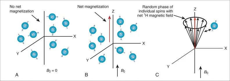
FIG. 1.1 Orientation of molecular spins. (A) With no magnetic field, the spins are oriented randomly, producing no net magnetization. (B) The spins orient with an applied magnetic field (B0), producing a net magnetic field aligned along B0. (C) The magnetized spins rotate around the B0 field (the Z axis) in a random way, resulting in no net magnetization in the X-Y plane.

where B is the magnetic field strength experienced by the spin, which is approximately B0, but includes any variations as described below, v is the spin's precessional frequency in cycles/s or hertz (Hz), and γ (gamma) is the gyromagnetic ratio, which is a constant related to the mass and charge of the water proton, γ/2π = 42.58 × 10⁶ Hz Tesla for water protons. The precessional frequency for water protons at 1.5 Tesla (T; a common CMR field strength) is ν = 63.87 × 10⁶ Hz, or roughly 64 MHz (about the frequency of an FM radio station and one of the reasons the CMR environment must be shielded from FM radio waves). Thus, at 3 T, the precessional frequency is twice this, ν = 127.74 × 10⁶ Hz (Eq. 1.1).
The Larmor equation (see Eq. 1.1) looks simple, but it is the basis of CMR imaging. To determine the location of different magnetic spins, the magnetic field (B) is made to vary (linearly) with position using specialized coils of wire (called “gradient coils”) inside the magnet. This results in a precessional frequency (v) that depends on a spin's location in the scanner. By measuring the number of spins precessing at each location (i.e., frequency), a magnetic resonance (MR) image is created1 as will be discussed below.
Detection of the MRI Signal
Alignment With the Main Magnetic Field
How can the MRI signal from water spins be detected in order to image the sample? Remember, when a sample (i.e., a patient) is placed in the main magnetic field (i.e., inside the bore of the magnet), the hydrogen spins align with that field (see Fig. 1.1B) and begin to precess at the Larmor frequency. We call the direction of the main magnetic field, B0, the Z-axis direction or the longitudinal direction. Since the spins partially align with the B0 field, they create a net magnetic field along the Z axis (oriented parallel with B0). However, the spins precess or rotate around the Z axis in a random, incoherent fashion, resulting in no net magnetization in the X-Y plane (also called the transverse plane) (see Fig. 1.1C). Magnetization by the B0 field (i.e., Mz) is not enough to generate an MR signal. The MR signal is measured as the magnetization on the X-Y plane (called Mxy). The MR signal is detected by placing a receiver coil (loops of wire tuned to receive signals oscillating at the Larmor frequency) near the subject to detect any spin precession in the X-Y plane. A receiver coil, placed to detect the precession of spins in the X-Y plane, will not detect any signal if Mxy is zero (see Fig. 1.1C).
Radiofrequency Excitation
To create an MR signal, the water spins must be precessing in a coherent manner in the X-Y plane. To accomplish this task, another (less powerful) magnetic field, with a strength that oscillates at the Larmor frequency is applied perpendicular to the main magnetic field (Fig. 1.2A). This process is called radiofrequency (RF) excitation, and the field is called an RF pulse or a B1 field, since it uses a magnetic (B1) field rotating at a high (radio wave) frequency. The frequency of this perpendicular B1 field has to precisely match the Larmor frequency of the water to excite the spins in the presence of the stronger B0 field. (This concept is analogous to exciting a piano string with a tuning fork. Only the string with the same resonant frequency as the tuning fork will efficiently absorb the energy from the fork and resonate. In fact, magnetic resonance imaging gets its name from the resonant frequency used for RF excitation.) The excitation field (B1) has a strength of only a few gauss, compared to the B0 field, which has a strength of 15,000 gauss at 1.5 T (1 T = 10,000 gauss). In addition, the B1 field is only applied transiently (for milliseconds), long enough to temporarily deflect the spins into a plane perpendicular to B0. This effect on the net magnetic field is illustrated in Fig. 1.2A, which shows a B1 pulse that drives the magnetization completely into the transverse plane (X-Y plane). This is called a 90-degree pulse (or saturation pulse), referring to the angle through which the initial magnetization (Mz) moves relative to the Z axis. However, in practice, the B1 pulse can be used to flip the magnetization by any angle from 0 to 180 degrees. A small B1 pulse (<90 degrees) is called an alpha pulse, while a 180-degree pulse is called an inversion pulse. A 180-degree flip is achieved by applying the B1 field for twice as long or with tw...
Table of contents
- Cover image
- Title Page
- Table of Contents
- Common Abbreviations Used in the Text
- Copyright
- Dedication
- Contributors
- Foreword
- Preface
- Acknowledgments
- Braunwald's Heart Disease Family of Books
- Section I Basic Principles of Cardiovascular Magnetic Resonance
- Section II Ischemic Heart Disease
- Section III Functional Disease
- Section IV Right Ventricular and Congenital Heart Disease
- Section V Vascular/Pericardium
- Section VI Interventional
- Section VII Economics and Guidelines
- Appendix A CMR Screening Form: Beth Israel Deaconess Medical Center (BIDMC)—CMR Center
- Appendix B CMR Sequence Protocols in Use (2018) at the Beth Israel Deaconess Medical Center (BIDMC)—CMR Center
- Appendix C Analogous CMR Terminology Used by Various Vendors
- Index
Frequently asked questions
Yes, you can cancel anytime from the Subscription tab in your account settings on the Perlego website. Your subscription will stay active until the end of your current billing period. Learn how to cancel your subscription
No, books cannot be downloaded as external files, such as PDFs, for use outside of Perlego. However, you can download books within the Perlego app for offline reading on mobile or tablet. Learn how to download books offline
Perlego offers two plans: Essential and Complete
- Essential is ideal for learners and professionals who enjoy exploring a wide range of subjects. Access the Essential Library with 800,000+ trusted titles and best-sellers across business, personal growth, and the humanities. Includes unlimited reading time and Standard Read Aloud voice.
- Complete: Perfect for advanced learners and researchers needing full, unrestricted access. Unlock 1.5M+ books across hundreds of subjects, including academic and specialized titles. The Complete Plan also includes advanced features like Premium Read Aloud and Research Assistant.
We are an online textbook subscription service, where you can get access to an entire online library for less than the price of a single book per month. With over 1.5 million books across 990+ topics, we’ve got you covered! Learn about our mission
Look out for the read-aloud symbol on your next book to see if you can listen to it. The read-aloud tool reads text aloud for you, highlighting the text as it is being read. You can pause it, speed it up and slow it down. Learn more about Read Aloud
Yes! You can use the Perlego app on both iOS and Android devices to read anytime, anywhere — even offline. Perfect for commutes or when you’re on the go.
Please note we cannot support devices running on iOS 13 and Android 7 or earlier. Learn more about using the app
Please note we cannot support devices running on iOS 13 and Android 7 or earlier. Learn more about using the app
Yes, you can access Cardiovascular Magnetic Resonance by Warren J. Manning,Dudley J. Pennell in PDF and/or ePUB format, as well as other popular books in Medicine & Cardiology. We have over 1.5 million books available in our catalogue for you to explore.