eBook - ePub
Cardiovascular Magnetic Resonance
A Companion to Braunwald's Heart Disease E-Book
Warren J. Manning, Dudley J. Pennell
This is a test
- 672 Seiten
- English
- ePUB (handyfreundlich)
- Über iOS und Android verfügbar
eBook - ePub
Cardiovascular Magnetic Resonance
A Companion to Braunwald's Heart Disease E-Book
Warren J. Manning, Dudley J. Pennell
Angaben zum Buch
Buchvorschau
Inhaltsverzeichnis
Quellenangaben
Über dieses Buch
- Provides state-of-the-art coverage of CMR technologies and guidelines, including basic principles, imaging techniques, ischemic heart disease, right ventricular and congenital heart disease, vascular and pericardium conditions, and functional cardiovascular disease.
- Includes new chapters on non-cardiac pathology, pacemaker safety, economics of CMR, and guidelines as well as new coverage of myocarditis and its diagnosis and assessment of prognosis by cardiovascular magnetic resonance, and the use of PET/CMR imaging of the heart, especially in sarcoidosis.
- Features more than 1, 100 high-quality images representing today's CMR imaging.
- Covers T1, T2 and ECV mapping, as well as T2* imaging in iron overload, which has been shown to save lives in patients with thalassaemia major
- Discusses the cost-effectiveness of CMR.
Häufig gestellte Fragen
Wie kann ich mein Abo kündigen?
Gehe einfach zum Kontobereich in den Einstellungen und klicke auf „Abo kündigen“ – ganz einfach. Nachdem du gekündigt hast, bleibt deine Mitgliedschaft für den verbleibenden Abozeitraum, den du bereits bezahlt hast, aktiv. Mehr Informationen hier.
(Wie) Kann ich Bücher herunterladen?
Derzeit stehen all unsere auf Mobilgeräte reagierenden ePub-Bücher zum Download über die App zur Verfügung. Die meisten unserer PDFs stehen ebenfalls zum Download bereit; wir arbeiten daran, auch die übrigen PDFs zum Download anzubieten, bei denen dies aktuell noch nicht möglich ist. Weitere Informationen hier.
Welcher Unterschied besteht bei den Preisen zwischen den Aboplänen?
Mit beiden Aboplänen erhältst du vollen Zugang zur Bibliothek und allen Funktionen von Perlego. Die einzigen Unterschiede bestehen im Preis und dem Abozeitraum: Mit dem Jahresabo sparst du auf 12 Monate gerechnet im Vergleich zum Monatsabo rund 30 %.
Was ist Perlego?
Wir sind ein Online-Abodienst für Lehrbücher, bei dem du für weniger als den Preis eines einzelnen Buches pro Monat Zugang zu einer ganzen Online-Bibliothek erhältst. Mit über 1 Million Büchern zu über 1.000 verschiedenen Themen haben wir bestimmt alles, was du brauchst! Weitere Informationen hier.
Unterstützt Perlego Text-zu-Sprache?
Achte auf das Symbol zum Vorlesen in deinem nächsten Buch, um zu sehen, ob du es dir auch anhören kannst. Bei diesem Tool wird dir Text laut vorgelesen, wobei der Text beim Vorlesen auch grafisch hervorgehoben wird. Du kannst das Vorlesen jederzeit anhalten, beschleunigen und verlangsamen. Weitere Informationen hier.
Ist Cardiovascular Magnetic Resonance als Online-PDF/ePub verfügbar?
Ja, du hast Zugang zu Cardiovascular Magnetic Resonance von Warren J. Manning, Dudley J. Pennell im PDF- und/oder ePub-Format sowie zu anderen beliebten Büchern aus Médecine & Cardiologie. Aus unserem Katalog stehen dir über 1 Million Bücher zur Verfügung.
Information
Section I
Basic Principles of Cardiovascular Magnetic Resonance
1
Basic Principles of Cardiovascular Magnetic Resonance
Robert S. Balaban, Dana C. Peters
Introduction
This introduction to the basic principles of cardiovascular magnetic resonance (CMR) describes the concepts of magnetization, T1, T2, T2*, and image formation, and describes some common CMR pulse sequences and parameters. These basic ideas will require some time and rereading to fully appreciate, but each new concept will add to the understanding of CMR.
The human body is composed of mostly water, and a lot of fat. The water (H2O) and fat contain many hydrogen atoms. Hydrogen atoms in turn are made up of a proton (1H, the hydrogen nucleus) and an electron. Hydrogen protons are in very high concentration in the body, roughly ~100 molar.a These hydrogen protons within the body can be imaged through magnetic resonance imaging (MRI). MRI is possible because hydrogen protons have “spin” and all nuclei with spin interact with magnetic fields. Spin is called the “intrinsic angular momentum,” and it is a fundamental property of protons. MRI physics is often called “spin physics” because it describes how the proton spins are used to image the body. In this chapter we will use the term spin to indicate the hydrogen proton's spin. MRI images these spins, and in this way it images the tissues in which they are embedded.
In the absence of a magnetic field, the spins are randomly oriented (Fig. 1.1A). But if placed in a large magnetic field (called B0; i.e., the CMR scanner), the water spins partly align in the same orientation of this applied magnetic field much like iron filings (Fig. 1.1B), with larger magnetic fields causing greater alignment of the spins. In fact, the bulk magnetization, Mz, is proportional to the strength of the applied field. Unlike iron filings, however, the interaction of the spins with the B0 field results in the spins rotating around the axis of the magnetic field (Fig. 1.1C). This is why the hydrogen nuclei in MRI are also referred to as spins: they spin (precess) around the B0 field. The frequency of precession (v) is a very important property of the spins in a magnetic field and is defined by the Larmor equation:
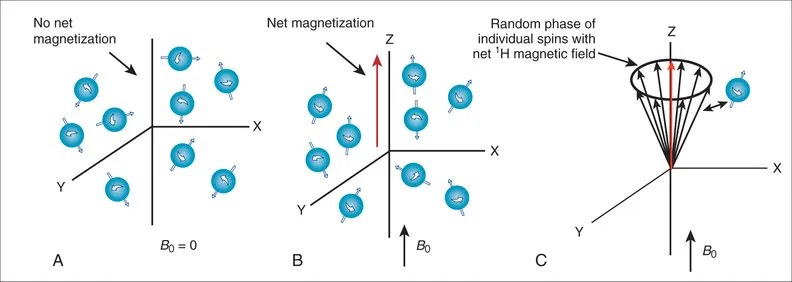
FIG. 1.1 Orientation of molecular spins. (A) With no magnetic field, the spins are oriented randomly, producing no net magnetization. (B) The spins orient with an applied magnetic field (B0), producing a net magnetic field aligned along B0. (C) The magnetized spins rotate around the B0 field (the Z axis) in a random way, resulting in no net magnetization in the X-Y plane.

where B is the magnetic field strength experienced by the spin, which is approximately B0, but includes any variations as described below, v is the spin's precessional frequency in cycles/s or hertz (Hz), and γ (gamma) is the gyromagnetic ratio, which is a constant related to the mass and charge of the water proton, γ/2π = 42.58 × 10⁶ Hz Tesla for water protons. The precessional frequency for water protons at 1.5 Tesla (T; a common CMR field strength) is ν = 63.87 × 10⁶ Hz, or roughly 64 MHz (about the frequency of an FM radio station and one of the reasons the CMR environment must be shielded from FM radio waves). Thus, at 3 T, the precessional frequency is twice this, ν = 127.74 × 10⁶ Hz (Eq. 1.1).
The Larmor equation (see Eq. 1.1) looks simple, but it is the basis of CMR imaging. To determine the location of different magnetic spins, the magnetic field (B) is made to vary (linearly) with position using specialized coils of wire (called “gradient coils”) inside the magnet. This results in a precessional frequency (v) that depends on a spin's location in the scanner. By measuring the number of spins precessing at each location (i.e., frequency), a magnetic resonance (MR) image is created1 as will be discussed below.
Detection of the MRI Signal
Alignment With the Main Magnetic Field
How can the MRI signal from water spins be detected in order to image the sample? Remember, when a sample (i.e., a patient) is placed in the main magnetic field (i.e., inside the bore of the magnet), the hydrogen spins align with that field (see Fig. 1.1B) and begin to precess at the Larmor frequency. We call the direction of the main magnetic field, B0, the Z-axis direction or the longitudinal direction. Since the spins partially align with the B0 field, they create a net magnetic field along the Z axis (oriented parallel with B0). However, the spins precess or rotate around the Z axis in a random, incoherent fashion, resulting in no net magnetization in the X-Y plane (also called the transverse plane) (see Fig. 1.1C). Magnetization by the B0 field (i.e., Mz) is not enough to generate an MR signal. The MR signal is measured as the magnetization on the X-Y plane (called Mxy). The MR signal is detected by placing a receiver coil (loops of wire tuned to receive signals oscillating at the Larmor frequency) near the subject to detect any spin precession in the X-Y plane. A receiver coil, placed to detect the precession of spins in the X-Y plane, will not detect any signal if Mxy is zero (see Fig. 1.1C).
Radiofrequency Excitation
To create an MR signal, the water spins must be precessing in a coherent manner in the X-Y plane. To accomplish this task, another (less powerful) magnetic field, with a strength that oscillates at the Larmor frequency is applied perpendicular to the main magnetic field (Fig. 1.2A). This process is called radiofrequency (RF) excitation, and the field is called an RF pulse or a B1 field, since it uses a magnetic (B1) field rotating at a high (radio wave) frequency. The frequency of this perpendicular B1 field has to precisely match the Larmor frequency of the water to excite the spins in the presence of the stronger B0 field. (This concept is analogous to exciting a piano string with a tuning fork. Only the string with the same resonant frequency as the tuning fork will efficiently absorb the energy from the fork and resonate. In fact, magnetic resonance imaging gets its name from the resonant frequency used for RF excitation.) The excitation field (B1) has a strength of only a few gauss, compared to the B0 field, which has a strength of 15,000 gauss at 1.5 T (1 T = 10,000 gauss). In addition, the B1 field is only applied transiently (for milliseconds), long enough to temporarily deflect the spins into a plane perpendicular to B0. This effect on the net magnetic field is illustrated in Fig. 1.2A, which shows a B1 pulse that drives the magnetization completely into the transverse plane (X-Y plane). This is called a 90-degree pulse (or saturation pulse), referring to the angle through which the initial magnetization (Mz) moves relative to the Z axis. However, in practice, the B1 pulse can be used to flip the magnetization by any angle from 0 to 180 degrees. A small B1 pulse (<90 degrees) is called an alpha pulse, while a 180-degree pulse is called an inversion pulse. A 180-degree flip is achieved by applying the B1 field for twice as long or with tw...
Inhaltsverzeichnis
Zitierstile für Cardiovascular Magnetic Resonance
APA 6 Citation
Manning, W., & Pennell, D. (2018). Cardiovascular Magnetic Resonance (3rd ed.). Elsevier Health Sciences. Retrieved from https://www.perlego.com/book/2938191/cardiovascular-magnetic-resonance-a-companion-to-braunwalds-heart-disease-ebook-pdf (Original work published 2018)
Chicago Citation
Manning, Warren, and Dudley Pennell. (2018) 2018. Cardiovascular Magnetic Resonance. 3rd ed. Elsevier Health Sciences. https://www.perlego.com/book/2938191/cardiovascular-magnetic-resonance-a-companion-to-braunwalds-heart-disease-ebook-pdf.
Harvard Citation
Manning, W. and Pennell, D. (2018) Cardiovascular Magnetic Resonance. 3rd edn. Elsevier Health Sciences. Available at: https://www.perlego.com/book/2938191/cardiovascular-magnetic-resonance-a-companion-to-braunwalds-heart-disease-ebook-pdf (Accessed: 15 October 2022).
MLA 7 Citation
Manning, Warren, and Dudley Pennell. Cardiovascular Magnetic Resonance. 3rd ed. Elsevier Health Sciences, 2018. Web. 15 Oct. 2022.