Principally based on dissections of hundreds of un-embalmed human cadavers over the past decade, Functional Atlas of the Human Fascial System presents a new vision of the human fascial system using anatomical and histological photographs along with microscopic analysis and biomechanical evaluation.Prof. Carla Stecco – orthopaedic surgeon and professor of anatomy and sport activities – brings together the research of a multi-specialist team of researchers and clinicians consisting of anatomists, biomechanical engineers, physiotherapists, osteopaths and plastic surgeons. In this Atlas Prof. Stecco presents for the first time a global view of fasciae and the actual connections that describe the myofascial kinetic chains. These descriptions help to explain how fascia plays a part in myofascial dysfunction and disease as well as how it may alter muscle function and disturb proprioceptive input. Prof. Stecco also highlights the continuity of the fascial planes, explaining the function of the fasciae and their connection between muscles, nerves and blood vessels. This understanding will help guide the practitioner in selecting the proper technique for a specific fascial problem with a view to enhancing manual therapy methods.Functional Atlas of the Human Fascial System opens with the first chapter classifying connective tissue and explaining its composition in terms of percentages of fibres, cells and extracellular matrix. The second chapter goes on to describe the general characteristics of the superficial fascia from a macroscopic and microscopic point of view; while the third analyzes the deep fascia in the same manner. The subsequent five chapters describe the fasciae from a topographical perspective. In this part of the Atlas, common anatomical terminology is used throughout to refer to the various fasciae but it also stresses the continuity of fasciae between the different bodily regions.- Over 300 unique photographs which show fascia on fresh (not embalmed) cadavers- Demonstrates the composition, form and function of the fascial system- Highlights the role of the deep fascia for proprioception and peripheral motor coordination- Companion website – www.atlasfascial.com – with videos showing how fascia connects with ligaments
- 256 pages
- English
- ePUB (mobile friendly)
- Available on iOS & Android
eBook - ePub
Functional Atlas of the Human Fascial System
About this book
Trusted by 375,005 students
Access to over 1.5 million titles for a fair monthly price.
Study more efficiently using our study tools.
Information
Topic
Medicine1
Connective Tissues
Composition of the Connective Tissues
Connective tissue (CT) is one of the four major classes of tissue (the others being epithelial, muscle and nerve tissue). It maintains the form of the body and its organs, and provides cohesion and structural support for the tissues and organs. CT derives its name from its function in connecting or binding cells and tissues. It is ubiquitous in the body and can be considered the ‘glue’ that holds the body parts together.
CT has three main components: cells, fibres and extracellular matrix (ECM) (Fig. 1.1). The cells provide the metabolic properties of the tissue, the fibres the mechanical properties, and the ECM the plasticity and malleability of the tissue. The most common cell types are fibroblasts, which produce collagen fibres and other intercellular materials. Cells such as adipocytes and undifferentiated mesenchymal cells are also present. The proportions of these three components vary from one part of the body to another depending on the local structural requirements. In some areas, the CT is loosely organized and highly cellular; in others, its fibrous components predominate; and in still others, the ground substance may be its most conspicuous feature. The consistency of the ECM is highly variable and ranges from gelatin like to a more rigid material. Consequently, the CT ranges in consistency from the gel-like softness of loose CT to the hardness of bone. The anatomical classification of the various types of CT is based largely upon the relative abundance and arrangement of these components. For example, strong CTs, such as tendons and ligaments, require a greater proportion of collagen fibres and fewer cells. Whereas, a CT composed mostly of cells like adipose tissue (fat) would not be very strong.
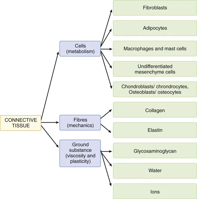
FIGURE 1.1 Composition of the CTs.
The CT has many functions:
• Structural support: it provides a structural framework for the body and maintains the anatomical form of the organs and systems. It forms the skeleton and the capsules surrounding the organs.
• Connection of body tissues: such as ligaments, tendons and fasciae.
• Protection of organs: it cushions and envelops the organs and separates them from surrounding structures. It permits necessary motilities between organs and fills the spaces between organs, preventing friction, pressure and damaging collisions among the mobile structures.
• Metabolical functions: provides a nutritive role. All the metabolites from the blood pass from capillary beds and diffuse through the adjacent CT to cells and tissues. Similarly waste metabolites from the cells and tissues diffuse through the loose CT before returning to the blood capillaries. The CT mediates and controls the various exchanges.
• Storage of energy: in the adipose tissue (a specialized CT).
• Regulation of diffusion of substances.
• Formation of scar tissue: it has a fundamental role in the recovery of the tissues following traumatic damage.
All CT cells are derived from mesenchymal cells. Mesenchymal cells are found in embryos and are, for the most part, derived from the middle germ layer of the embryo (mesoderm), but some of the CTs of the head region are derived from the neural crest (ectodermal in origin). Mesenchymal cells are only found in embryos, although some mesenchyme-like cells persist in adult CT and retain their capacity to differentiate in response to injury.
Extracellular Matrix
The ECM refers to the extracellular components of CT and supporting tissues. This matrix distributes the mechanical stresses on tissues and provides the structural environment for the cells embedded in it. It forms a framework to which cells adhere and on which they can move (Standring 2008), and is composed of a ground substance and fibres. The ground substance is composed of water, extracellular proteins, glycosaminoglycans (GAGs) and proteoglycans, in varying proportions. It is clear, colourless and viscous. The fibres are of different types, but the principal ones are collagen and elastin and these define the mechanical properties of the tissue.
Ground Substance
Ground substance is an amorphous gel-like substance surrounding the cells. It does not include fibres (e.g. collagen and elastic fibres), but does include all the other components of the ECM and is also known as the extrafibrillar matrix. The ground substance is responsible for the support and nutrition of cells. It determines the compliance, mobility and integrity of the CT and is a lubricant and binder for the diverse elements of the ECM (Hukinsa & Aspden 1985). The presence of macromolecules in the ground substance allows the collagen fibres to slide with little friction, when force is applied, providing relative mobility until the interfibrillar cross-links are tensioned. Both collagens and water molecules have electric conductive and polarization properties, as do the matrix molecules. Polarization waves are possible and protons can ‘jump’ along the collagen fibres much faster than electrical signals conducted by nerves (Jaroszyk & Marzec 1993).
Proteoglycans
The ground substance contains proteoglycans that are very large macromolecules consisting of a core protein to which many GAG molecules are covalently attached, somewhat like the bristles around the stem of a bottle brush. GAGs are long-chained polysaccharides made up of repeating disaccharide units and one of the sugars in each disaccharide unit is a glycosamine, hence the name GAG. Many of the sugars in GAGs have sulfate and carboxyl groups, which makes them highly negatively charged. A family of seven distinct GAGs is recognized, based on differences in the specific sugar residues, nature of the linkages and degree of sulfation. These GAGs are hyaluronan, chondroitin-4-sulfate, chondroitin-6-sulfate, dermatan sulfate, keratin sulfate, heparin sulfate and heparin.
GAGs are not flexible enough to assume a globular form and stay extended occupying an ample surface in relationship to their volume. The high density of negative charges attracts water forming a hydrated gel; this gel is responsible for the turgidity and viscoelasticity of the CT and controls the diffusion of various metabolites. In particular, it permits the rapid diffusion of water-soluble molecules but inhibits the movement of large molecules and bacteria. Its viscoelasticity allows the tissue to return to its original form after stress, and enables the collagen fibres to move without friction against each other, to absorb forces that affect the tissue and to protect the collagen network from excessive stress. The various levels of water determine if the ground matrix is a sol or gel and subsequently the mobility of the collagen fibres that are embedded inside. Smaller proteoglycans such as decorin, which has a single GAG chain, could play a role in the organization and disposition of collagen fibres. Proteoglycans are also found in cellular membranes and inside cells, and mediate the interactions of the cells and ground substance.
Hyaluronan
Hyaluronan (HA) is the GAG most represented in loose CT and is the only one that has no sulfate groups. It differs from the typical GAG because it is extremely long and rigid: consisting of a chain of several thousand sugars, as compared to several hundred or less found in other GAGs. It also does not bind to a core protein to become part of a proteoglycan; instead, proteoglycans indirectly bind to HA via special linker proteins to form giant macromolecules. These hydrophilic macromolecules are particularly abundant in cartilage ground substance and are responsible for the turgor pressure that gives cartilage its shape. HA provides the structure as well as turgor to the aqueous of the eye and protects fetal vessels from compression in the Wharton's jelly of the umbilical cord (Fig. 1.2).
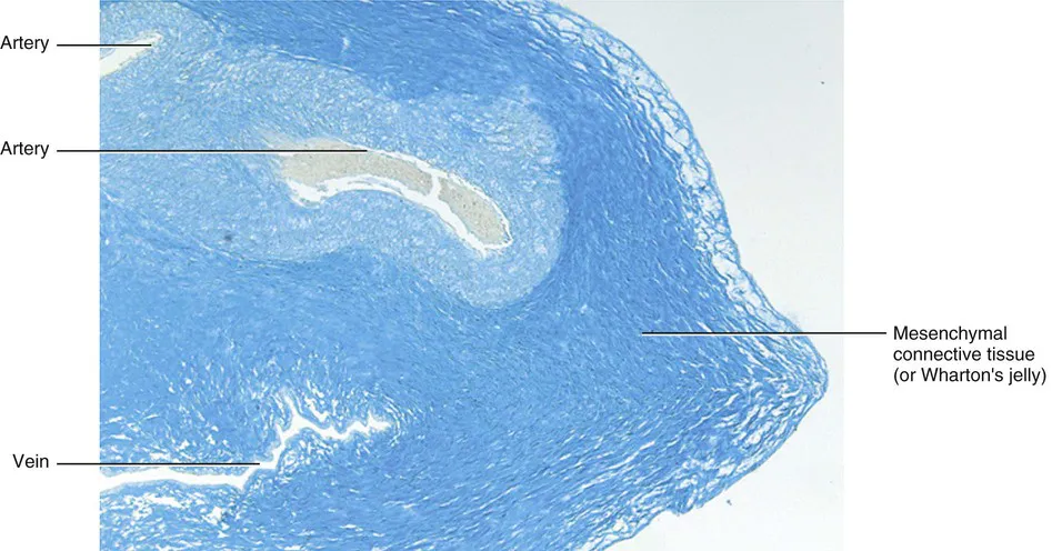
FIGURE 1.2 Histology of the umbilical cord, Alcian blue, enlargement 50×. Note how much the mesenchymal CT stains blue, highlighting the abundance of hyaluronan in the ECM of the umbilical cord.
HA provides moisture for the skin by means of its large volume of solvent water (as much as 10,000 times the volume of the original material). It also provides a lubricant for muscles and tendons as they glide over skeletal or under aponeurotic fasciae. It is likely that these gliding interactions are influenced by the composition and efficacy of the HA-rich ECM. This HA-rich layer also protects muscles and supports recovery from injury, and stimulates satellite cell proliferation following loss of muscle fibres. Changes in this HA-rich matrix contribute to pain, inflammation and loss of function. HA is abundant during the earliest stages of wound healing and functions to open up tissue spaces through which cells can travel. By binding to cell receptors and interacting with the cytoskeleton it confers motility to the cells.
HA is particularly plentiful during embryogenesis and in tissues undergoing rapid growth, and is present wherever repair and regeneration occur. Depending on its chain length, especially when it becomes fragmented, HA has recently been shown to have a wide range of opposing biological functions: such as becoming angiogenic, inflammatory and immunostimulatory.
The turnover of HA is about ...
Table of contents
- Cover image
- Title page
- Table of Contents
- Copyright
- Foreword
- Preface
- Acknowledgements
- 1 Connective Tissues
- 2 Subcutaneous Tissue and Superficial Fascia
- 3 Deep Fasciae
- 4 Fasciae of the Head and Neck
- 5 Fasciae of the Thorax and Abdomen
- 6 Fasciae of the Back
- 7 Fasciae of the Upper Limb
- 8 Fasciae of the Lower Limb
- Index
Frequently asked questions
Yes, you can cancel anytime from the Subscription tab in your account settings on the Perlego website. Your subscription will stay active until the end of your current billing period. Learn how to cancel your subscription
No, books cannot be downloaded as external files, such as PDFs, for use outside of Perlego. However, you can download books within the Perlego app for offline reading on mobile or tablet. Learn how to download books offline
Perlego offers two plans: Essential and Complete
- Essential is ideal for learners and professionals who enjoy exploring a wide range of subjects. Access the Essential Library with 800,000+ trusted titles and best-sellers across business, personal growth, and the humanities. Includes unlimited reading time and Standard Read Aloud voice.
- Complete: Perfect for advanced learners and researchers needing full, unrestricted access. Unlock 1.5M+ books across hundreds of subjects, including academic and specialized titles. The Complete Plan also includes advanced features like Premium Read Aloud and Research Assistant.
We are an online textbook subscription service, where you can get access to an entire online library for less than the price of a single book per month. With over 1.5 million books across 990+ topics, we’ve got you covered! Learn about our mission
Look out for the read-aloud symbol on your next book to see if you can listen to it. The read-aloud tool reads text aloud for you, highlighting the text as it is being read. You can pause it, speed it up and slow it down. Learn more about Read Aloud
Yes! You can use the Perlego app on both iOS and Android devices to read anytime, anywhere — even offline. Perfect for commutes or when you’re on the go.
Please note we cannot support devices running on iOS 13 and Android 7 or earlier. Learn more about using the app
Please note we cannot support devices running on iOS 13 and Android 7 or earlier. Learn more about using the app
Yes, you can access Functional Atlas of the Human Fascial System by Carla Stecco, Warren I Hammer in PDF and/or ePUB format, as well as other popular books in Medicine & Alternative & Complementary Medicine. We have over 1.5 million books available in our catalogue for you to explore.