In this unique book, Dr. Bertorini guides you through more than 100 cases that demonstrate the diagnosis and management of a wide range of common and rare neuromuscular disorders. No other reference boasts such a large array of clinical studies devoted to all areas of this broad topic! Each case study reviews the etiologies, pathogenesis, differential diagnosis, and management of a particular disorder, helping you not only recognize its presentation, but also determine a diagnosis and the best treatment plans for your patients. You'll also find expert guidance on the basic mechanisms of neuromuscular disorders, clinical examination, and diagnostic tests—including EMG, muscle biopsy, genetic testing, and more.- More than 100 detailed case studies explore both common and rare neuromuscular disorders and the treatment protocols for each, equipping you with the knowledge you need to confidently manage any challenge. Each case includes a summary of important points or highlights of the study.- Case studies are arranged either by complaint or by diagnosis so that you can successfully manage your patients with or without an initial diagnosis.- Comprehensive coverage of EMGs and nerve conduction studies and other diagnostic tests, including muscle and nerve biopsies and genetic testing, helps you accurately diagnose nerve, muscle, and neuromuscular transmission disorders.- Detailed discussions of treatment plans and commonly used drugs enhance your management of autoimmune disorders, painful neuropathy, dysautonomia, and other neuromuscular disorders.- A reader-friendly format takes you step by step through the diagnosis and treatment of neuromuscular disorders, from the basic anatomy and physiology of the nerve and muscle through to clinical evaluation, diagnostic testing, and therapy.- More than 350 high-quality illustrations, including full-color patient photographs, biopsies, and EMG tracings, make complex concepts easier to understand and apply.
- 632 pages
- English
- ePUB (mobile friendly)
- Available on iOS & Android
eBook - ePub
Neuromuscular Case Studies
About this book
Trusted by 375,005 students
Access to over 1.5 million titles for a fair monthly price.
Study more efficiently using our study tools.
Information
1 Neuromuscular Anatomy and Function
Neuromuscular disorders are those that affect the anterior horn (motor neuron diseases, such as amyotrophic lateral sclerosis [ALS]), roots (radiculopathies), plexuses (plexopathies), or peripheral nerves (polyneuropathy, mononeuropathy). These also include diseases of the neuromuscular junction (e.g., myasthenia gravis) and muscle fibers (myopathies) (Fig. 1-1). Several of these disorders could involve other regions of the nervous system such as the spinal cord and its pathways, or other organs in the body. The following is a review of the basic anatomy and physiology of muscle and nerve that is of importance in understanding these disorders.
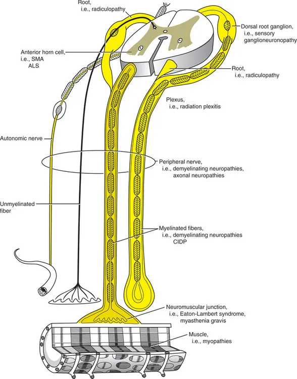
FIGURE 1-1 The anatomic elements of the peripheral nervous system. Related neurologic disorders are in parentheses.
(Reprinted with permission from Bertorini TE: Overview and classification of neuromuscular disorders. In Bertorini TE [ed]: Clinical Evaluation and Diagnostic Tests for Neuromuscular Disorders. Boston, Butterworth-Heinemann, 2002.)
The performance of movements requires the interaction of neuronal systems of the cerebral cortex and the motor neurons of the brainstem and anterior horns of the spinal cord. The fine modulation of these movements is regulated by several pathways that include the proprioceptive input for feedback, the interaction of the cortical neurons, limbic system, brainstem, and interneuronal systems.1
The feedback is regulated by the interaction of receptors in muscle spindles and deep tendon organs. The muscle spindle has intrafusal muscle fibers that when stretched activate their 1 alpha nerve fibers which stimulate motor neurons of agonist muscles to contract. They also activate inhibitory neurons that go to motor neurons of antagonist muscles. The sensitivity of the spindles varies with their length, which is determined by the contraction of its intrafusal fibers that are innervated by gamma motor neurons. Another regulatory mechanism is the input of the deep tendon organs through their 1b afferent axons which are activated upon changes in muscle tension, causing the inhibition of agonist motor neurons, while facilitating antagonist muscles to contract.
The motor unit is the final pathway of the motor system. This is formed by the motor neurons of the spinal cord or brainstem, their myelinated axons, and the muscle fibers innervated by that neuron, which are intermixed with fibers from other motor units. The physiologic and biochemical characteristics of muscle fibers of a motor unit are determined by the rate of firing of their motor neurons.2
There are two major types of muscle fibers, depending on their speed of contraction, their biochemical characteristics, and, thus, their histochemical staining, and all muscle fibers of a motor unit are of the same type. Type I fibers correspond to the red or dark meat in animals. Type II muscle fibers correspond to white meat (Table 1-1). The characteristics of these fibers could be changed by cross innervation from nerves of one type of muscle to the other or by prolonged stimulation of their axons at different rates. Type I muscle fibers are slow contracting and stain pale with ATPase using alkaline pH, and dark with oxidative stains.1,2 These fibers also have subtypes that can be recognized by special histochemical stains, such as, for example, non-specific esterase and menadione-mediated alpha glycerophosphate dehydrogenase.3
Table 1-1. Major Skeletal Muscle Fiber Types
| Type I | Type II | |
|---|---|---|
| Contraction time | Slow (tonic) | Fast (twitch) |
| Oxidative enzyme content (i.e., NADH-TR*) | High | Low |
| Capillary supply | Rich | Poor |
| Myofibrillar adenosine triphosphatase (pH 9.4) | Low | High |
| Myofibrillar adenosine triphosphatase (pH 4.3) | High | Low |
| Glycolytic activity | Low | High |
| Lipid content | High | Low |
NADH-TR, nicotinamide adenine dinucleotide-tetrazolium reductase.
Reprinted from Bertorini TE (ed): Clinical Evaluation and Diagnostic Tests for Neuromuscular Disorders. Boston, Butterworth-Heinemann, 2002, p 600.
Type II fibers stain dark with ATPase at alkaline pH and have two major subtypes. Type IIA are fast-contracting, nonfatigable fibers that stain dark with alkaline ATPase and pale at acid pH of 4.6 and 4.3. Their function depends mainly on aerobic metabolism. The type IIB fibers stain intermediate with ATPase at pH 4.6 (Fig. 1-2). These are fast-contracting, fast-fatiguing fibers that depend mainly on glycolytic, anaerobic metabolism.2
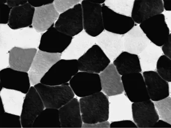
FIGURE 1-2 Muscle biopsy stained with ATPase at pH of 4.6. Notice the dark type I fibers, pale type IIA fibers, and intermediate type 2B fibers (×200).
In humans, muscle fibers of both fiber types and subtypes are intermixed with fibers of other motor units. They appear in muscle histology in an almost checkerboard pattern, as seen in Figure 1-2, with predominance of one or the other in some muscles. The deltoid, for example, has mainly type I fibers, and the quadriceps has mainly type II.
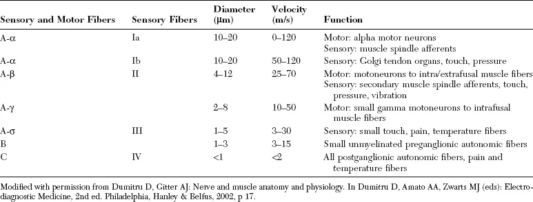
Peripheral nerves carry axons from motor neurons and sensory afferents from the Golgi’s tendon organs and spindles. They also contain large myelinated fibers that carry proprioceptive sensation. Nerves also have unmyelinated and small myelinated axons that carry touch, pain, and temperature sensations. Their cell bodies are located in the dorsal root ganglia; peripheral nerves also have autonomic fibers with myelinated presynaptic and unmyelinated postsynaptic axons (Table 1-2).
Table 1-2. Nerve Fiber Classification
ANATOMY OF THE CRANIAL AND PERIPHERAL NERVES
Human striated muscles are innervated by nerves that originate in the brainstem and spinal cord.4-7 These are summarized here. Motor cranial nerves include those to the extraocular muscles such as the oculomotor, abducens, and trochlear nerves; and the V cranial or trigeminal nerve which innervates muscles of mastication and provides sensation to the face. The facial, or VII cranial nerve innervates muscles of facial expression, as well as the lacrimal and salivary glands, provides sensation and taste to the anterior part of the tongue, and relays sensation of the tympanic membrane, external auditory canal, and ...
Table of contents
- Cover
- Title Page
- Copyright
- Dedication
- Preface
- Acknowledgments
- Table of Contents
- Chapter 1: Neuromuscular Anatomy and Function
- Chapter 2: Neurologic Evaluation and Ancillary Tests
- Chapter 3: Therapy in Neuromuscular Diseases
- CASE 1: An Elderly Woman with Numbness in the Hands
- CASE 2: A Diabetic Man with Arm Swelling, Pain, and Numbness
- CASE 3: A Diabetic Man with Hand Numbness and Fainting Spells
- CASE 4: A Man with Hand Pain
- CASE 5: A Woman with Hand Weakness, Numbness, and Pain After Cardiac Catheterization
- CASE 6: A Man Who Practices Karate and Has Forearm Pain
- CASE 7: A Man with a History of Polio Who Developed Hand Weakness and Numbness
- CASE 8: A Young Woman with Multiple Sclerosis and Hand Numbness
- CASE 9: A Woman with a Wrist Drop and a Mass in the Arm
- CASE 10: A Man with Bilateral Arm Weakness After Lumbar Surgery
- CASE 11: A Woman with Acute Onset of Shoulder Pain and Weakness
- CASE 12: A Man with Lymphoma and Numbness in the Fingers of One Hand
- CASE 13: A Woman with Arm Weakness After Treatment for Breast Cancer
- CASE 14: A Diabetic Man with Shoulder Pain and Weakness
- CASE 15: A Woman with Arm Weakness and a Skin Rash
- CASE 16: A Woman with Neck and Arm Pain
- CASE 17: A Woman with Neck Pain and a Weak Arm
- CASE 18: A Woman with Back Pain and Leg Weakness
- CASE 19: A Woman with Back Pain and Weakness
- CASE 20: A Woman with Back Pain and Unusual Nerve Conduction Tests
- CASE 21: A Man with Pheochromocytoma and Back Pain
- CASE 22: A Diabetic Man with Back and Leg Pain
- CASE 23: A Man with Leg Pain While Walking
- CASE 24: A Man with Foot Pain
- CASE 25: A Man with Unilateral Proximal Leg Weakness After Heart Catheterization
- CASE 26: A Woman with Uterine Cancer and Unilateral Leg Weakness
- CASE 27: A Man with Burning Sensations in the Thigh
- CASE 28: A Man with Weakness and Fasciculations in the Lower Extremities
- CASE 29: A Woman with Unilateral Facial Weakness
- CASE 30: A Diabetic Woman with Muscle Weakness and Ophthalmoplegia
- CASE 31: A Man with a Dropped Head
- CASE 32: A Man in the “Barrel”
- CASE 33: A Man with a Long History of Muscle Weakness in One Arm
- CASE 34: A Man with Progressive Leg Muscle Weakness 20 Years After an Acute Paralysis
- CASE 35: A Man with One Large Calf
- CASE 36: A Man with Muscle Spasms and Elevated Serum Creatine Kinase
- CASE 37: A Man with Muscle Twitching and Poor Balance
- CASE 38: A Floppy Child
- CASE 39: A Man with Acute Areflexic Paralysis and Central Nervous System Symptoms
- CASE 40: A Man with Acute Muscle Weakness and Respiratory Failure
- CASE 41: A Young Woman with Acute Onset of Clumsiness and Ophthalmoplegia
- CASE 42: A Woman with Seafood Poisoning in Catfish Country
- CASE 43: A Woman with a Kingly Disease
- CASE 44: A Myasthenic Woman with Persistent Weakness
- CASE 45: A Man with Neck Pain and Numbness in the Hands
- CASE 46: A Woman with Peripheral Neuropathy and Frequent Falls
- CASE 47: A Man with Weakness in the Legs
- CASE 48: An Elderly Woman with Asymmetric Weakness and a Monoclonal Gammopathy
- CASE 49: A Man with Progressive Weakness and Fasciculations
- CASE 50: A Man with Diabetic Polyneuropathy Who Developed Rapidly Progressive Weakness
- CASE 51: An Older Man with Leg Numbness
- CASE 52: A Woman with a Neuropathy and Symptoms of a Central Nervous System Disease
- CASE 53: A Man with Progressive Weakness After a Gastroplasty
- CASE 54: A Diabetic Woman with Proximal Leg Weakness and Pain
- CASE 55: A Diabetic Man with Bilateral Arm Weakness
- CASE 56: A Diabetic Man with Pain and Swelling in the Thigh
- CASE 57: A Uremic Man with Burning Feet
- CASE 58A: A Woman with Leg Numbness, Pain, Weakness, and Slow Nerve Conduction Velocities
- CASE 58B: A Boy with Leg Weakness
- CASE 59: A Man with Hand Weakness and Numbness After Skiing
- CASE 60: A Man with Recurrent Foot Drop
- CASE 61: An Elderly Woman with Foot Drop and Hand Weakness
- CASE 62: A Woman with Rheumatoid Arthritis Who Developed Numbness and Pain in the Feet
- CASE 63: A Woman with Granulomatous Lesions in the Lungs and a Peripheral Neuropathy
- CASE 64: A Man with a Neuropathy, Weight Loss, and Lung Nodules
- CASE 65: A Woman with Difficulty Walking and Ataxia
- CASE 66: A Man with Progressive Neuropathy and Congestive Heart Failure
- CASE 67: An Elderly Woman with a Progressive Neuropathy
- CASE 68: A Young Woman with Difficulty Swallowing
- CASE 69: A Young Woman with Intermittent Weakness and a Positive Family History of Similar Problems
- CASE 70: A Woman with Muscle Weakness and Areflexia
- CASE 71: An Elderly Woman with Proximal Weakness
- CASE 72: A Young Woman with Distal Upper Extremity Weakness
- CASE 73A: A Young Man with Episodic Weakness
- CASE 73B: An African American Man with Hyperthyroidism and Acute Paralysis
- CASE 74: A Boy with Large Muscles, Leg Pain, and Elevated Serum Creatine Kinase
- CASE 75: A Woman with Proximal Muscle Weakness, Calf Hypertrophy, Heel Contractures, and Elevated Serum Creatine Kinase
- CASE 76: A Young Man with Distal Leg Wasting and Normal Sensation
- CASE 77: A Woman with Droopy Eyelids and Difficulty Swallowing
- CASE 78: A Man with Facial and Shoulder Muscle Weakness
- CASE 79: A Boy Born Floppy with Severe Weakness, and Later, Contractures
- CASE 80: A Woman with Long-standing Weakness and Foot Deformities
- CASE 81: A Woman with Droopy Eyelids and Ophthalmoplegia
- CASE 82: A Man with Weakness and Swelling in the Neck
- CASE 83: A Woman with Muscle Pains and Dark Urine
- CASE 84: A Woman with Progressive Proximal Muscle Weakness
- CASE 85: A Sedentary Man with Acute Respiratory Failure and Myoglobinuria
- CASE 86: A Uremic Man with Proximal Muscle Weakness
- CASE 87: A Woman with Weakness, Elevated Cholesterol, and Serum Creatine Kinase Levels
- CASE 88: An Older Woman with Leg Weakness and Atrophic Muscle Fibers on Biopsy
- CASE 89A: A Woman with Muscle Weakness and a Skin Rash
- CASE 89B: A Child with Muscle Weakness and a Skin Rash
- CASE 90: A Man with Muscle Pains
- CASE 91: An Older Woman with Progressive Muscle Weakness
- CASE 92: An HIV-Infected Man with Muscle Weakness and Spasms
- CASE 93: A Woman with Limb Swelling and Pain
- CASE 94: A Man with a Bent Spine
- CASE 95: A Man with Muscle Stiffness, and Later with Diplopia
- CASE 96: A Woman with Muscle Cramps and Fasciculations
- CASE 97: A Man with Large and Stiff Muscles
- CASE 98: A Boy with Short Stature, Small Jaw, Muscle Hypertrophy, and Stiffness
- CASE 99: An Elderly Woman with Muscle Spasms
- CASE 100: A Woman with Proximal Muscle Weakness and Neuromuscular Irritability
- CASE 101: A Woman with Focal Spontaneous Muscle Movements
- Index of Cases by Diagnosis
- Index
Frequently asked questions
Yes, you can cancel anytime from the Subscription tab in your account settings on the Perlego website. Your subscription will stay active until the end of your current billing period. Learn how to cancel your subscription
No, books cannot be downloaded as external files, such as PDFs, for use outside of Perlego. However, you can download books within the Perlego app for offline reading on mobile or tablet. Learn how to download books offline
Perlego offers two plans: Essential and Complete
- Essential is ideal for learners and professionals who enjoy exploring a wide range of subjects. Access the Essential Library with 800,000+ trusted titles and best-sellers across business, personal growth, and the humanities. Includes unlimited reading time and Standard Read Aloud voice.
- Complete: Perfect for advanced learners and researchers needing full, unrestricted access. Unlock 1.5M+ books across hundreds of subjects, including academic and specialized titles. The Complete Plan also includes advanced features like Premium Read Aloud and Research Assistant.
We are an online textbook subscription service, where you can get access to an entire online library for less than the price of a single book per month. With over 1.5 million books across 990+ topics, we’ve got you covered! Learn about our mission
Look out for the read-aloud symbol on your next book to see if you can listen to it. The read-aloud tool reads text aloud for you, highlighting the text as it is being read. You can pause it, speed it up and slow it down. Learn more about Read Aloud
Yes! You can use the Perlego app on both iOS and Android devices to read anytime, anywhere — even offline. Perfect for commutes or when you’re on the go.
Please note we cannot support devices running on iOS 13 and Android 7 or earlier. Learn more about using the app
Please note we cannot support devices running on iOS 13 and Android 7 or earlier. Learn more about using the app
Yes, you can access Neuromuscular Case Studies by Tulio E. Bertorini in PDF and/or ePUB format, as well as other popular books in Medicine & Neurology. We have over 1.5 million books available in our catalogue for you to explore.