
eBook - ePub
Neuromuscular Case Studies
Tulio E. Bertorini
This is a test
Buch teilen
- 632 Seiten
- English
- ePUB (handyfreundlich)
- Über iOS und Android verfügbar
eBook - ePub
Neuromuscular Case Studies
Tulio E. Bertorini
Angaben zum Buch
Buchvorschau
Inhaltsverzeichnis
Quellenangaben
Über dieses Buch
In this unique book, Dr. Bertorini guides you through more than 100 cases that demonstrate the diagnosis and management of a wide range of common and rare neuromuscular disorders. No other reference boasts such a large array of clinical studies devoted to all areas of this broad topic! Each case study reviews the etiologies, pathogenesis, differential diagnosis, and management of a particular disorder, helping you not only recognize its presentation, but also determine a diagnosis and the best treatment plans for your patients. You'll also find expert guidance on the basic mechanisms of neuromuscular disorders, clinical examination, and diagnostic tests—including EMG, muscle biopsy, genetic testing, and more.
- More than 100 detailed case studies explore both common and rare neuromuscular disorders and the treatment protocols for each, equipping you with the knowledge you need to confidently manage any challenge. Each case includes a summary of important points or highlights of the study.
- Case studies are arranged either by complaint or by diagnosis so that you can successfully manage your patients with or without an initial diagnosis.
- Comprehensive coverage of EMGs and nerve conduction studies and other diagnostic tests, including muscle and nerve biopsies and genetic testing, helps you accurately diagnose nerve, muscle, and neuromuscular transmission disorders.
- Detailed discussions of treatment plans and commonly used drugs enhance your management of autoimmune disorders, painful neuropathy, dysautonomia, and other neuromuscular disorders.
- A reader-friendly format takes you step by step through the diagnosis and treatment of neuromuscular disorders, from the basic anatomy and physiology of the nerve and muscle through to clinical evaluation, diagnostic testing, and therapy.
- More than 350 high-quality illustrations, including full-color patient photographs, biopsies, and EMG tracings, make complex concepts easier to understand and apply.
Häufig gestellte Fragen
Wie kann ich mein Abo kündigen?
Gehe einfach zum Kontobereich in den Einstellungen und klicke auf „Abo kündigen“ – ganz einfach. Nachdem du gekündigt hast, bleibt deine Mitgliedschaft für den verbleibenden Abozeitraum, den du bereits bezahlt hast, aktiv. Mehr Informationen hier.
(Wie) Kann ich Bücher herunterladen?
Derzeit stehen all unsere auf Mobilgeräte reagierenden ePub-Bücher zum Download über die App zur Verfügung. Die meisten unserer PDFs stehen ebenfalls zum Download bereit; wir arbeiten daran, auch die übrigen PDFs zum Download anzubieten, bei denen dies aktuell noch nicht möglich ist. Weitere Informationen hier.
Welcher Unterschied besteht bei den Preisen zwischen den Aboplänen?
Mit beiden Aboplänen erhältst du vollen Zugang zur Bibliothek und allen Funktionen von Perlego. Die einzigen Unterschiede bestehen im Preis und dem Abozeitraum: Mit dem Jahresabo sparst du auf 12 Monate gerechnet im Vergleich zum Monatsabo rund 30 %.
Was ist Perlego?
Wir sind ein Online-Abodienst für Lehrbücher, bei dem du für weniger als den Preis eines einzelnen Buches pro Monat Zugang zu einer ganzen Online-Bibliothek erhältst. Mit über 1 Million Büchern zu über 1.000 verschiedenen Themen haben wir bestimmt alles, was du brauchst! Weitere Informationen hier.
Unterstützt Perlego Text-zu-Sprache?
Achte auf das Symbol zum Vorlesen in deinem nächsten Buch, um zu sehen, ob du es dir auch anhören kannst. Bei diesem Tool wird dir Text laut vorgelesen, wobei der Text beim Vorlesen auch grafisch hervorgehoben wird. Du kannst das Vorlesen jederzeit anhalten, beschleunigen und verlangsamen. Weitere Informationen hier.
Ist Neuromuscular Case Studies als Online-PDF/ePub verfügbar?
Ja, du hast Zugang zu Neuromuscular Case Studies von Tulio E. Bertorini im PDF- und/oder ePub-Format sowie zu anderen beliebten Büchern aus Médecine & Neurologie. Aus unserem Katalog stehen dir über 1 Million Bücher zur Verfügung.
Information
Thema
MédecineThema
Neurologie1 Neuromuscular Anatomy and Function
Neuromuscular disorders are those that affect the anterior horn (motor neuron diseases, such as amyotrophic lateral sclerosis [ALS]), roots (radiculopathies), plexuses (plexopathies), or peripheral nerves (polyneuropathy, mononeuropathy). These also include diseases of the neuromuscular junction (e.g., myasthenia gravis) and muscle fibers (myopathies) (Fig. 1-1). Several of these disorders could involve other regions of the nervous system such as the spinal cord and its pathways, or other organs in the body. The following is a review of the basic anatomy and physiology of muscle and nerve that is of importance in understanding these disorders.
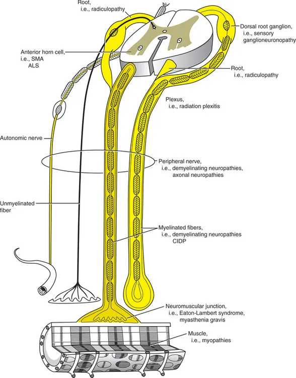
FIGURE 1-1 The anatomic elements of the peripheral nervous system. Related neurologic disorders are in parentheses.
(Reprinted with permission from Bertorini TE: Overview and classification of neuromuscular disorders. In Bertorini TE [ed]: Clinical Evaluation and Diagnostic Tests for Neuromuscular Disorders. Boston, Butterworth-Heinemann, 2002.)
The performance of movements requires the interaction of neuronal systems of the cerebral cortex and the motor neurons of the brainstem and anterior horns of the spinal cord. The fine modulation of these movements is regulated by several pathways that include the proprioceptive input for feedback, the interaction of the cortical neurons, limbic system, brainstem, and interneuronal systems.1
The feedback is regulated by the interaction of receptors in muscle spindles and deep tendon organs. The muscle spindle has intrafusal muscle fibers that when stretched activate their 1 alpha nerve fibers which stimulate motor neurons of agonist muscles to contract. They also activate inhibitory neurons that go to motor neurons of antagonist muscles. The sensitivity of the spindles varies with their length, which is determined by the contraction of its intrafusal fibers that are innervated by gamma motor neurons. Another regulatory mechanism is the input of the deep tendon organs through their 1b afferent axons which are activated upon changes in muscle tension, causing the inhibition of agonist motor neurons, while facilitating antagonist muscles to contract.
The motor unit is the final pathway of the motor system. This is formed by the motor neurons of the spinal cord or brainstem, their myelinated axons, and the muscle fibers innervated by that neuron, which are intermixed with fibers from other motor units. The physiologic and biochemical characteristics of muscle fibers of a motor unit are determined by the rate of firing of their motor neurons.2
There are two major types of muscle fibers, depending on their speed of contraction, their biochemical characteristics, and, thus, their histochemical staining, and all muscle fibers of a motor unit are of the same type. Type I fibers correspond to the red or dark meat in animals. Type II muscle fibers correspond to white meat (Table 1-1). The characteristics of these fibers could be changed by cross innervation from nerves of one type of muscle to the other or by prolonged stimulation of their axons at different rates. Type I muscle fibers are slow contracting and stain pale with ATPase using alkaline pH, and dark with oxidative stains.1,2 These fibers also have subtypes that can be recognized by special histochemical stains, such as, for example, non-specific esterase and menadione-mediated alpha glycerophosphate dehydrogenase.3
Table 1-1. Major Skeletal Muscle Fiber Types
| Type I | Type II | |
|---|---|---|
| Contraction time | Slow (tonic) | Fast (twitch) |
| Oxidative enzyme content (i.e., NADH-TR*) | High | Low |
| Capillary supply | Rich | Poor |
| Myofibrillar adenosine triphosphatase (pH 9.4) | Low | High |
| Myofibrillar adenosine triphosphatase (pH 4.3) | High | Low |
| Glycolytic activity | Low | High |
| Lipid content | High | Low |
NADH-TR, nicotinamide adenine dinucleotide-tetrazolium reductase.
Reprinted from Bertorini TE (ed): Clinical Evaluation and Diagnostic Tests for Neuromuscular Disorders. Boston, Butterworth-Heinemann, 2002, p 600.
Type II fibers stain dark with ATPase at alkaline pH and have two major subtypes. Type IIA are fast-contracting, nonfatigable fibers that stain dark with alkaline ATPase and pale at acid pH of 4.6 and 4.3. Their function depends mainly on aerobic metabolism. The type IIB fibers stain intermediate with ATPase at pH 4.6 (Fig. 1-2). These are fast-contracting, fast-fatiguing fibers that depend mainly on glycolytic, anaerobic metabolism.2
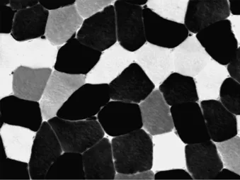
FIGURE 1-2 Muscle biopsy stained with ATPase at pH of 4.6. Notice the dark type I fibers, pale type IIA fibers, and intermediate type 2B fibers (×200).
In humans, muscle fibers of both fiber types and subtypes are intermixed with fibers of other motor units. They appear in muscle histology in an almost checkerboard pattern, as seen in Figure 1-2, with predominance of one or the other in some muscles. The deltoid, for example, has mainly type I fibers, and the quadriceps has mainly type II.
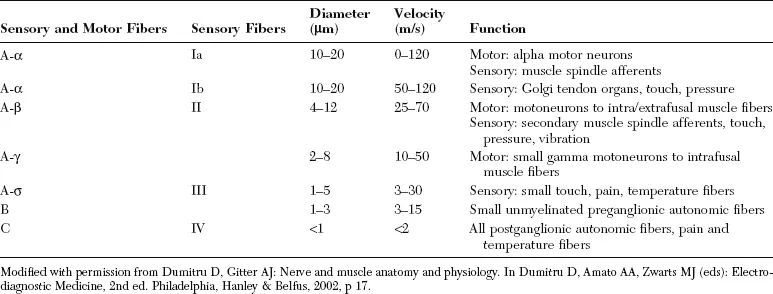
Peripheral nerves carry axons from motor neurons and sensory afferents from the Golgi’s tendon organs and spindles. They also contain large myelinated fibers that carry proprioceptive sensation. Nerves also have unmyelinated and small myelinated axons that carry touch, pain, and temperature sensations. Their cell bodies are located in the dorsal root ganglia; peripheral nerves also have autonomic fibers with myelinated presynaptic and unmyelinated postsynaptic axons (Table 1-2).
Table 1-2. Nerve Fiber Classification
ANATOMY OF THE CRANIAL AND PERIPHERAL NERVES
Human striated muscles are innervated by nerves that originate in the brainstem and spinal cord.4-7 These are summarized here. Motor cranial nerves include those to the extraocular muscles such as the oculomotor, abducens, and trochlear nerves; and the V cranial or trigeminal nerve which innervates muscles of mastication and provides sensation to the face. The facial, or VII cranial nerve innervates muscles of facial expression, as well as the lacrimal and salivary glands, provides sensation and taste to the anterior part of the tongue, and relays sensation of the tympanic membrane, external auditory canal, and ...