Taking the place of the multiple texts traditionally needed to cover visual anatomy and physiology, Clinical Anatomy and Physiology of the Visual System, 3rd Edition dramatically lightens your load by providing one book that covers it all! This concise, well-referenced resource contains information on the clinical anatomy of the eye, its adnexa and visual pathways, histologic information, plus newly added content on physiology of the human ocular structures. Vivid illustrations complement the text and provide clinical information on diseases and disorders that represent departures from normal clinical anatomy.- Comprehensive physiology coverage clarifies the integration between structure and function, eliminating your need for multiple books on the anatomy and physiology of the visual system.- An emphasis on clinical application helps you better understand the processes that occur in disease and dysfunction.- Genetic information keeps you current with the latest developments in visual anatomy and physiology.- Full-color illustrations throughout the text enhance your understanding of anatomical and clinical information.- UNIQUE! Clinical Comment sections provide a solid foundation for recognizing and understanding clinical situations, conditions, diseases, and treatments.- Photos of normal eye structures illustrate clinical appearance and demonstrate how appearance is directly related to structure.- Geriatric coverage, including aging changes in ocular tissue and the visual pathway, keeps you up-to-date with the expanding field of geriatric care.- UNIQUE! Expert coverage written by an actual optometrist gives you a practical framework for recognizing and understanding clinical situations, problems, and treatments.

eBook - ePub
Clinical Anatomy of the Visual System E-Book
Clinical Anatomy of the Visual System E-Book
- 302 pages
- English
- ePUB (mobile friendly)
- Available on iOS & Android
eBook - ePub
Clinical Anatomy of the Visual System E-Book
Clinical Anatomy of the Visual System E-Book
About this book
Trusted by 375,005 students
Access to over 1.5 million titles for a fair monthly price.
Study more efficiently using our study tools.
Information
Topic
MedicineSubtopic
Opthalmology & OptometryChapter 1 Visual System
The visual system takes in information from the environment in the form of light and analyzes and interprets it. This process of sight and visual perception involves a complex system of structures, each of which is designed for a specific purpose. The organization of each structure enables it to perform its intended function.
The eye houses the elements that take in light rays and changes them to a neural signal; it is protected by its location within the bone and connective tissue framework of the orbit. The eyelids cover and protect the anterior surface of the eye and contain glands that produce the lubricating tear film. Muscles, attached to the outer coat of the eye, control and direct the globe’s movement, and the muscles of both eyes are coordinated to provide binocular vision. A network of blood vessels supplies nutrients, and a complex system of nerves provides sensory and motor innervation to the eye and surrounding tissues and structures. The neural signal that carries visual information passes through a complex and intricately designed pathway within the central nervous system, enabling an accurate view of the surrounding environment. This information, evaluated by a process called visual perception, influences myriad decisions and activities.
This book examines the macroscopic and microscopic anatomy and physiology of the components in this complex system and the structures that support it.
The Eye
Anatomic Features
The eye is a special sense organ made up of three coats, or tunics, as follows:
1. The outer fibrous layer of connective tissue forms the cornea and sclera.
2. The middle vascular layer is composed of the iris, ciliary body, and choroid.
3. The inner neural layer is the retina.
Within this globe are three spaces: the anterior chamber, posterior chamber, and vitreous chamber. The crystalline lens is located in the region of the posterior chamber (Figure 1-1).
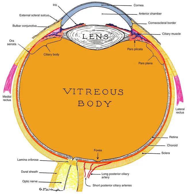
Figure 1-1 The visual system.
(From Kronfeld PC: The human eye, Rochester, NY, 1943, Bausch & Lomb Press.)
The outer dense connective tissue of the eye provides protection for the structures within and maintains the shape of the globe, providing resistance to the pressure of the fluids inside. The sclera is the opaque white of the eye and is covered by the transparent conjunctiva. The transparent cornea allows light rays to enter the globe and, by refraction, helps bring these light rays into focus on the retina. The region in which the transition from cornea to sclera and conjunctiva occurs is the limbus.
The vascular layer of the eye is the uvea, which is made up of three structures, each having a separate function but all are interconnected. Some of the histologic layers are continuous throughout all three structures and are derived from the same embryonic germ cell layer. The iris is the most anterior structure, acting as a diaphragm to regulate the amount of light entering the pupil. The two iris muscles control the shape and diameter of the pupil and are supplied by the autonomic nervous system. Continuous with the iris at its root is the ciliary body, which produces the components of the aqueous humor and contains the muscle that controls the shape of the lens. The posterior part of the uvea, the choroid, is an anastomosing network of blood vessels with a dense capillary network; it surrounds the retina and supplies nutrients to the outer retinal layers.
The neural tissue of the retina, by complex biochemical processes, changes light energy into a signal that can be transmitted along a neural pathway. The signal passes through the retina, exits the eye through the optic nerve, and is transmitted to various parts of the brain for processing.
The interior of the eye is made up of three chambers. The anterior chamber is bounded in front by the cornea and posteriorly by the iris and anterior surface of the lens. The posterior chamber lies behind the iris and surrounds the equator of the lens, separating it from the ciliary body. The anterior and posterior chambers are continuous with one another through the pupil, and both contain aqueous humor that is produced by the ciliary body. The aqueous humor provides nourishment for the surrounding structures, particularly the cornea and lens. The vitreous chamber, which is the largest space, lies adjacent to the inner retinal layer and is bounded in front by the lens. This chamber contains a gel-like substance, the vitreous humor.
The crystalline lens is located in the area of the posterior chamber and provides additional refractive power for accurately focusing images onto the retina. The lens must change shape to view an object that is close to the eye, through the mechanism of accommodation.
Anatomic Directions and Planes
Anatomy is an exacting science, and specific terminology is basic to its discussion. The following anatomic directions should be familiar (Figure 1-2):
• Anterior, or ventral: toward the front
• Posterior, or dorsal: toward the back
• Superior, or cranial: toward the head
• Inferior, or caudal: away from the head
• Medial: toward the midline
• Lateral: away from the midline
• Proximal: near the point of origin
• Distal: away from the point of origin
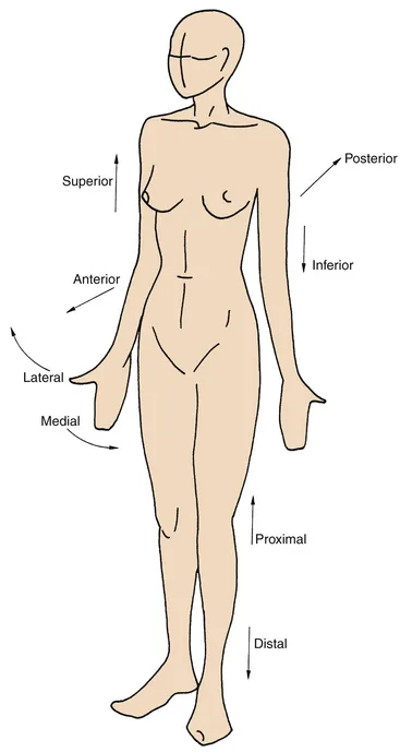
Figure 1-2 Anatomic directions.
(From Palastanga N, Field D, Soames R: Anatomy and human movement, Oxford, UK, 1989, Butterworth-Heinemann.)
The following planes are used in describing anatomic structures (Figure 1-3):
• Sagittal: vertical plane running from anterior to posterior locations, dividing the structure into right and left sides.
• Midsagittal: sagittal plane through the midline, dividing the structure into right and left halves.
• Coronal or frontal: vertical plane running from side to side, dividing the structure into anterior and posterior parts.
• Transverse: horizontal plane dividing the structure into superior and inferior parts.
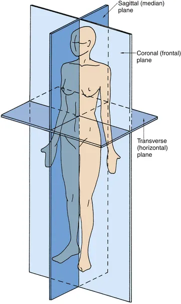
Figure 1-3 Anatomic planes.
(From Palastanga N, Field D, Soames R: Anatomy and human movement, Oxford, UK, 1989, Butterworth-Heinemann.)
Because the globe is a spherical structure, references to locations can sometimes be confusing. In references to anterior and posterior locations of the globe, the anterior pole (i.e., center of the cornea) is the reference point. For example, the pupil is anterior to the ciliary body (see Figure 1-1). When layers or structures are referred to as inner or outer, the reference is to the entire globe unless specified otherwise. The point of reference is the center of the globe, which would lie within the vitreous. For example, the retina is inner to the sclera (see Figure 1-1). In addition, the term sclerad is used to mean “toward the sclera,” and vitread is used to mean “toward the vitreous.”
Refractive Conditions
If the refractive power of the optical components of the eye, primarily the cornea and lens, correlate with the distances between the cornea, lens, and retina so that incoming parallel light rays come into focus on the retina, a clear image will be seen. This condition is called emmetropia (Figure 1-4, A). No correction is necessary for clear distance vision. In hyperopia (farsightedness), the distance from the cornea to the retina is too short for the refractive power of the cornea and lens, thereby causing images that would come into focus behind the retina (Figure 1-4, B). Hyperopia can be corrected by placing a convex lens in front of the eye to increase the convergence of the incoming light rays. In myopia (nearsightedness), because the lens and cornea are too strong or, more likely, the eyeball is too long, parallel light rays are brought into focus in front of the retina (Figure 1-4, C). Myopia can be corrected by plac...
Table of contents
- Cover
- Title Page
- Front Matter
- Copyright
- Dedication
- Preface
- Acknowledgments
- Table of Contents
- Chapter 1: Visual System
- Chapter 2: Cornea and Sclera
- Chapter 3: Uvea
- Chapter 4: Retina
- Chapter 5: Crystalline Lens
- Chapter 6: Aqueous and Vitreous Humors
- Chapter 7: Ocular Embryology
- Chapter 8: Bones of the Skull and Orbit
- Chapter 9: Ocular Adnexa and Lacrimal System
- Chapter 10: Extraocular Muscles
- Chapter 11: Orbital Blood Supply
- Chapter 12: Cranial Nerve Innervation of Ocular Structures
- Chapter 13: Visual Pathway
- Chapter 14: Autonomic Innervation of Ocular Structures
- Index
Frequently asked questions
Yes, you can cancel anytime from the Subscription tab in your account settings on the Perlego website. Your subscription will stay active until the end of your current billing period. Learn how to cancel your subscription
No, books cannot be downloaded as external files, such as PDFs, for use outside of Perlego. However, you can download books within the Perlego app for offline reading on mobile or tablet. Learn how to download books offline
Perlego offers two plans: Essential and Complete
- Essential is ideal for learners and professionals who enjoy exploring a wide range of subjects. Access the Essential Library with 800,000+ trusted titles and best-sellers across business, personal growth, and the humanities. Includes unlimited reading time and Standard Read Aloud voice.
- Complete: Perfect for advanced learners and researchers needing full, unrestricted access. Unlock 1.5M+ books across hundreds of subjects, including academic and specialized titles. The Complete Plan also includes advanced features like Premium Read Aloud and Research Assistant.
We are an online textbook subscription service, where you can get access to an entire online library for less than the price of a single book per month. With over 1.5 million books across 990+ topics, we’ve got you covered! Learn about our mission
Look out for the read-aloud symbol on your next book to see if you can listen to it. The read-aloud tool reads text aloud for you, highlighting the text as it is being read. You can pause it, speed it up and slow it down. Learn more about Read Aloud
Yes! You can use the Perlego app on both iOS and Android devices to read anytime, anywhere — even offline. Perfect for commutes or when you’re on the go.
Please note we cannot support devices running on iOS 13 and Android 7 or earlier. Learn more about using the app
Please note we cannot support devices running on iOS 13 and Android 7 or earlier. Learn more about using the app
Yes, you can access Clinical Anatomy of the Visual System E-Book by Lee Ann Remington,Denise Goodwin in PDF and/or ePUB format, as well as other popular books in Medicine & Opthalmology & Optometry. We have over 1.5 million books available in our catalogue for you to explore.