
eBook - ePub
Clinical Anatomy of the Visual System E-Book
Lee Ann Remington, Denise Goodwin
This is a test
Buch teilen
- 302 Seiten
- English
- ePUB (handyfreundlich)
- Über iOS und Android verfügbar
eBook - ePub
Clinical Anatomy of the Visual System E-Book
Lee Ann Remington, Denise Goodwin
Angaben zum Buch
Buchvorschau
Inhaltsverzeichnis
Quellenangaben
Über dieses Buch
Taking the place of the multiple texts traditionally needed to cover visual anatomy and physiology, Clinical Anatomy and Physiology of the Visual System, 3 rd Edition dramatically lightens your load by providing one book that covers it all! This concise, well-referenced resource contains information on the clinical anatomy of the eye, its adnexa and visual pathways, histologic information, plus newly added content on physiology of the human ocular structures. Vivid illustrations complement the text and provide clinical information on diseases and disorders that represent departures from normal clinical anatomy.
- Comprehensive physiology coverage clarifies the integration between structure and function, eliminating your need for multiple books on the anatomy and physiology of the visual system.
- An emphasis on clinical application helps you better understand the processes that occur in disease and dysfunction.
- Genetic information keeps you current with the latest developments in visual anatomy and physiology.
- Full-color illustrations throughout the text enhance your understanding of anatomical and clinical information.
- UNIQUE! Clinical Comment sections provide a solid foundation for recognizing and understanding clinical situations, conditions, diseases, and treatments.
- Photos of normal eye structures illustrate clinical appearance and demonstrate how appearance is directly related to structure.
- Geriatric coverage, including aging changes in ocular tissue and the visual pathway, keeps you up-to-date with the expanding field of geriatric care.
- UNIQUE! Expert coverage written by an actual optometrist gives you a practical framework for recognizing and understanding clinical situations, problems, and treatments.
Häufig gestellte Fragen
Wie kann ich mein Abo kündigen?
Gehe einfach zum Kontobereich in den Einstellungen und klicke auf „Abo kündigen“ – ganz einfach. Nachdem du gekündigt hast, bleibt deine Mitgliedschaft für den verbleibenden Abozeitraum, den du bereits bezahlt hast, aktiv. Mehr Informationen hier.
(Wie) Kann ich Bücher herunterladen?
Derzeit stehen all unsere auf Mobilgeräte reagierenden ePub-Bücher zum Download über die App zur Verfügung. Die meisten unserer PDFs stehen ebenfalls zum Download bereit; wir arbeiten daran, auch die übrigen PDFs zum Download anzubieten, bei denen dies aktuell noch nicht möglich ist. Weitere Informationen hier.
Welcher Unterschied besteht bei den Preisen zwischen den Aboplänen?
Mit beiden Aboplänen erhältst du vollen Zugang zur Bibliothek und allen Funktionen von Perlego. Die einzigen Unterschiede bestehen im Preis und dem Abozeitraum: Mit dem Jahresabo sparst du auf 12 Monate gerechnet im Vergleich zum Monatsabo rund 30 %.
Was ist Perlego?
Wir sind ein Online-Abodienst für Lehrbücher, bei dem du für weniger als den Preis eines einzelnen Buches pro Monat Zugang zu einer ganzen Online-Bibliothek erhältst. Mit über 1 Million Büchern zu über 1.000 verschiedenen Themen haben wir bestimmt alles, was du brauchst! Weitere Informationen hier.
Unterstützt Perlego Text-zu-Sprache?
Achte auf das Symbol zum Vorlesen in deinem nächsten Buch, um zu sehen, ob du es dir auch anhören kannst. Bei diesem Tool wird dir Text laut vorgelesen, wobei der Text beim Vorlesen auch grafisch hervorgehoben wird. Du kannst das Vorlesen jederzeit anhalten, beschleunigen und verlangsamen. Weitere Informationen hier.
Ist Clinical Anatomy of the Visual System E-Book als Online-PDF/ePub verfügbar?
Ja, du hast Zugang zu Clinical Anatomy of the Visual System E-Book von Lee Ann Remington, Denise Goodwin im PDF- und/oder ePub-Format sowie zu anderen beliebten Büchern aus Medicine & Opthalmology & Optometry. Aus unserem Katalog stehen dir über 1 Million Bücher zur Verfügung.
Information
Chapter 1 Visual System
The visual system takes in information from the environment in the form of light and analyzes and interprets it. This process of sight and visual perception involves a complex system of structures, each of which is designed for a specific purpose. The organization of each structure enables it to perform its intended function.
The eye houses the elements that take in light rays and changes them to a neural signal; it is protected by its location within the bone and connective tissue framework of the orbit. The eyelids cover and protect the anterior surface of the eye and contain glands that produce the lubricating tear film. Muscles, attached to the outer coat of the eye, control and direct the globe’s movement, and the muscles of both eyes are coordinated to provide binocular vision. A network of blood vessels supplies nutrients, and a complex system of nerves provides sensory and motor innervation to the eye and surrounding tissues and structures. The neural signal that carries visual information passes through a complex and intricately designed pathway within the central nervous system, enabling an accurate view of the surrounding environment. This information, evaluated by a process called visual perception, influences myriad decisions and activities.
This book examines the macroscopic and microscopic anatomy and physiology of the components in this complex system and the structures that support it.
The Eye
Anatomic Features
The eye is a special sense organ made up of three coats, or tunics, as follows:
1. The outer fibrous layer of connective tissue forms the cornea and sclera.
2. The middle vascular layer is composed of the iris, ciliary body, and choroid.
3. The inner neural layer is the retina.
Within this globe are three spaces: the anterior chamber, posterior chamber, and vitreous chamber. The crystalline lens is located in the region of the posterior chamber (Figure 1-1).
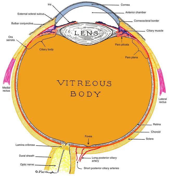
Figure 1-1 The visual system.
(From Kronfeld PC: The human eye, Rochester, NY, 1943, Bausch & Lomb Press.)
The outer dense connective tissue of the eye provides protection for the structures within and maintains the shape of the globe, providing resistance to the pressure of the fluids inside. The sclera is the opaque white of the eye and is covered by the transparent conjunctiva. The transparent cornea allows light rays to enter the globe and, by refraction, helps bring these light rays into focus on the retina. The region in which the transition from cornea to sclera and conjunctiva occurs is the limbus.
The vascular layer of the eye is the uvea, which is made up of three structures, each having a separate function but all are interconnected. Some of the histologic layers are continuous throughout all three structures and are derived from the same embryonic germ cell layer. The iris is the most anterior structure, acting as a diaphragm to regulate the amount of light entering the pupil. The two iris muscles control the shape and diameter of the pupil and are supplied by the autonomic nervous system. Continuous with the iris at its root is the ciliary body, which produces the components of the aqueous humor and contains the muscle that controls the shape of the lens. The posterior part of the uvea, the choroid, is an anastomosing network of blood vessels with a dense capillary network; it surrounds the retina and supplies nutrients to the outer retinal layers.
The neural tissue of the retina, by complex biochemical processes, changes light energy into a signal that can be transmitted along a neural pathway. The signal passes through the retina, exits the eye through the optic nerve, and is transmitted to various parts of the brain for processing.
The interior of the eye is made up of three chambers. The anterior chamber is bounded in front by the cornea and posteriorly by the iris and anterior surface of the lens. The posterior chamber lies behind the iris and surrounds the equator of the lens, separating it from the ciliary body. The anterior and posterior chambers are continuous with one another through the pupil, and both contain aqueous humor that is produced by the ciliary body. The aqueous humor provides nourishment for the surrounding structures, particularly the cornea and lens. The vitreous chamber, which is the largest space, lies adjacent to the inner retinal layer and is bounded in front by the lens. This chamber contains a gel-like substance, the vitreous humor.
The crystalline lens is located in the area of the posterior chamber and provides additional refractive power for accurately focusing images onto the retina. The lens must change shape to view an object that is close to the eye, through the mechanism of accommodation.
Anatomic Directions and Planes
Anatomy is an exacting science, and specific terminology is basic to its discussion. The following anatomic directions should be familiar (Figure 1-2):
• Anterior, or ventral: toward the front
• Posterior, or dorsal: toward the back
• Superior, or cranial: toward the head
• Inferior, or caudal: away from the head
• Medial: toward the midline
• Lateral: away from the midline
• Proximal: near the point of origin
• Distal: away from the point of origin
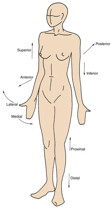
Figure 1-2 Anatomic directions.
(From Palastanga N, Field D, Soames R: Anatomy and human movement, Oxford, UK, 1989, Butterworth-Heinemann.)
The following planes are used in describing anatomic structures (Figure 1-3):
• Sagittal: vertical plane running from anterior to posterior locations, dividing the structure into right and left sides.
• Midsagittal: sagittal plane through the midline, dividing the structure into right and left halves.
• Coronal or frontal: vertical plane running from side to side, dividing the structure into anterior and posterior parts.
• Transverse: horizontal plane dividing the structure into superior and inferior parts.
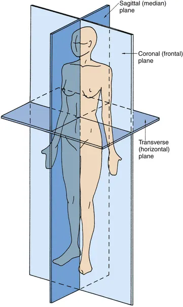
Figure 1-3 Anatomic planes.
(From Palastanga N, Field D, Soames R: Anatomy and human movement, Oxford, UK, 1989, Butterworth-Heinemann.)
Because the globe is a spherical structure, references to locations can sometimes be confusing. In references to anterior and posterior locations of the globe, the anterior pole (i.e., center of the cornea) is the reference point. For example, the pupil is anterior to the ciliary body (see Figure 1-1). When layers or structures are referred to as inner or outer, the reference is to the entire globe unless specified otherwise. The point of reference is the center of the globe, which would lie within the vitreous. For example, the retina is inner to the sclera (see Figure 1-1). In addition, the term sclerad is used to mean “toward the sclera,” and vitread is used to mean “toward the vitreous.”
Refractive Conditions
If the refractive power of the optical components of the eye, primarily the cornea and lens, correlate with the distances between the cornea, lens, and retina so that incoming parallel light rays come into focus on the retina, a clear image will be seen. This condition is called emmetropia (Figure 1-4, A). No correction is necessary for clear distance vision. In hyperopia (farsightedness), the distance from the cornea to the retina is too short for the refractive power of the cornea and lens, thereby causing images that would come into focus behind the retina (Figure 1-4, B). Hyperopia can be corrected by placing a convex lens in front of the eye to increase the convergence of the incoming light rays. In myopia (nearsightedness), because the lens and cornea are too strong or, more likely, the eyeball is too long, parallel light rays are brought into focus in front of the retina (Figure 1-4, C). Myopia can be corrected by plac...