Richly illustrated and comprehensive in scope, Obstetric Imaging, 2nd Edition, provides up-to-date, authoritative guidelines for more than 200 obstetric conditions and procedures, keeping you at the forefront of this fast-changing field. This highly regarded reference covers the extensive and ongoing advances in maternal and fetal imaging in a concise, newly streamlined format for quicker access to common and uncommon findings. Detailed, expert guidance, accompanied by superb, high-quality images, helps you make the most of new technologies and advances in obstetric imaging.- Features more than 1, 350 high-quality images, including 400 in color.- Helps you select the best imaging approaches and effectively interpret your findings with a highly templated, bulleted, at-a-glance organization.- Reflects all the latest developments in the field, including genetics, open fetal surgery, fetal echocardiography, Zika virus, and 3D imaging, so you can provide the safest and most responsive care to both mother and fetus.- Includes new chapters on Limbs and Bones Overview; Open Fetal Surgery; Biophysical Profile; Ultrasound Physics; Elastography; Doppler; MRI; Echogenic Bowel; Pregnancy of Unknown Location (PUL), Failed Pregnancy and Ectopic Pregnancy, Cesarean Scar Pregnancy; Cytomegalovirus (CMG), Rubella, Toxoplasmosis, Herpes, Varicella; and Congenital Syphilis; plus a new chapter on Zika Virus written by imaging experts from the "hot zone."- Keeps you up to date with the latest developments in multimodality imaging and optimizing diagnostic accuracy from ultrasound, 3D ultrasound, Doppler, MRI, elastography, image-guided interventions, and much more.- Expert Consult™ eBook version included with purchase. This enhanced eBook experience allows you to search all of the text, figures, Q&As, and references from the book on a variety of devices.
eBook - ePub
Obstetric Imaging: Fetal Diagnosis and Care E-Book
- 848 pages
- English
- ePUB (mobile friendly)
- Available on iOS & Android
eBook - ePub
Obstetric Imaging: Fetal Diagnosis and Care E-Book
About this book
Trusted by 375,005 students
Access to over 1.5 million titles for a fair monthly price.
Study more efficiently using our study tools.
Information
Topic
MedicinePart 1
Atlas of Selected Normal Images
1
Atlas of Selected Normal Images
Mert Ozan Bahtiyar, Carole Gravino
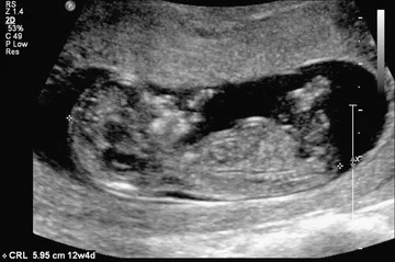
Fig. 1.1 The crown-rump length is a measurement of the length of human fetuses from the top of the crown to the bottom of the rump. It is used to estimate gestational age.
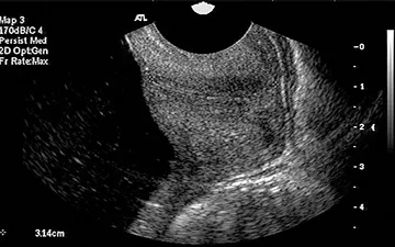
Fig. 1.2 Transvaginal ultrasound and sagittal long-axis view of the endocervical canal. Both the internal os and the external os are well visualized. The cervical length is measured from the internal os to the external os along the endocervical canal.
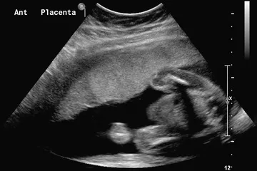
Fig. 1.3 Sagittal view of the uterus with an anterior (Ant) placenta.
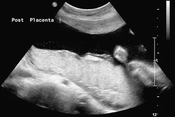
Fig. 1.4 Sagittal view of the uterus showing a posterior (Post) placenta.
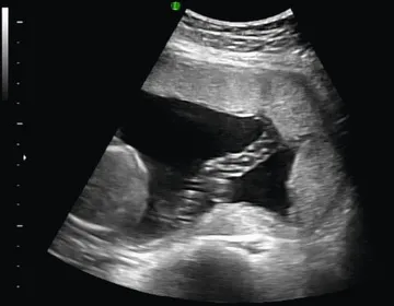
Fig. 1.5 Normal umbilical cord insertion into the placenta.
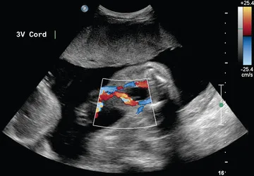
Fig. 1.6 Fetal umbilical cord insertion site. The umbilical arteries emerge caudally—originating at the iliac arteries and coursing along the margin of the urinary bladder. The umbilical vein proceeds cephalad and joins the fetal portal circulation. 3V, Three-vessel.
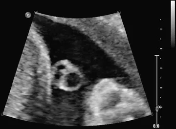
Fig. 1.7 Transverse view of the umbilical cord. The umbilical cord is composed of a vein and two smaller arteries.
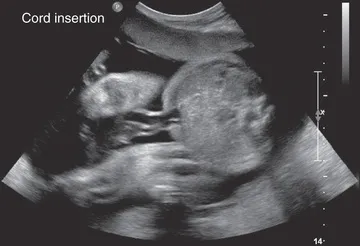
Fig. 1.8 Transverse view of the fetal abdomen and the umbilical cord insertion site showing integrity of the central abdominal wall.
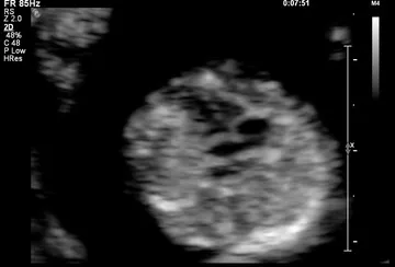
Fig. 1.9 Four-chamber view of the fetal heart at 12 weeks of gestation.
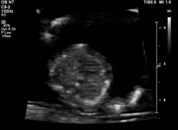
Fig. 1.10 Interventricular septum at 12 weeks of gestation.
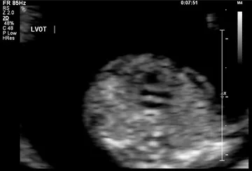
Fig. 1.11 Left ventricular outflow at 12 weeks of gestation. LVOT, Left ventricular outflow tract.
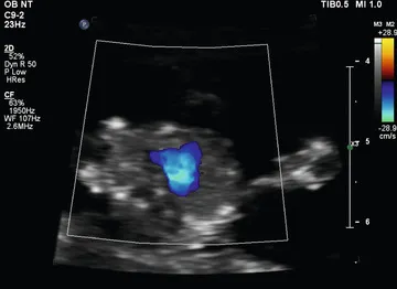
Fig. 1.12 Three-vessel view shown by color Doppler at 12 weeks of gestation.
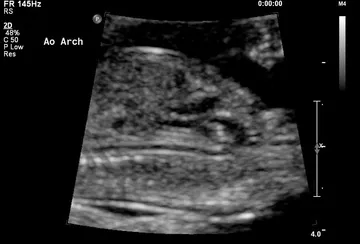
Fig. 1.13 Aortic arch (Ao Arch) at 12 weeks of gestation.
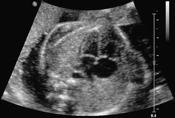
Fig. 1.14 Four-chamber view obtained with a transverse axial view through the fetal thorax. This view provides information on the size of the heart and its chambers; the pulmonary venous connections to the atrial segment; the morphology of the ventricles; the type of atrioventricular (AV) connection; and ...
Table of contents
- Cover image
- Title Page
- Table of Contents
- Copyright
- Contributors
- Preface
- Video Contents
- Part 1 Atlas of Selected Normal Images
- Part 2 Thorax
- Part 3 Retroperitoneum
- Part 4 Abdomen
- Part 5 Central Nervous System
- Part 6 First-Trimester Complications
- Part 7 Skeletal Dysplasias: An Overview
- Part 8 Head and Neck
- Part 9 Heart and Great Vessels
- Part 10 Placenta and Cord
- Part 11 Fetal Growth
- Part 12 Procedures
- Part 13 Miscellany
- Part 14 Syndromes
- Part 15 Chromosomes
- Part 16 Multiple Gestation
- Part 17 Infections
- Part 18 Technology and New Ultrasound Applications
- Index
Frequently asked questions
Yes, you can cancel anytime from the Subscription tab in your account settings on the Perlego website. Your subscription will stay active until the end of your current billing period. Learn how to cancel your subscription
No, books cannot be downloaded as external files, such as PDFs, for use outside of Perlego. However, you can download books within the Perlego app for offline reading on mobile or tablet. Learn how to download books offline
Perlego offers two plans: Essential and Complete
- Essential is ideal for learners and professionals who enjoy exploring a wide range of subjects. Access the Essential Library with 800,000+ trusted titles and best-sellers across business, personal growth, and the humanities. Includes unlimited reading time and Standard Read Aloud voice.
- Complete: Perfect for advanced learners and researchers needing full, unrestricted access. Unlock 1.5M+ books across hundreds of subjects, including academic and specialized titles. The Complete Plan also includes advanced features like Premium Read Aloud and Research Assistant.
We are an online textbook subscription service, where you can get access to an entire online library for less than the price of a single book per month. With over 1.5 million books across 990+ topics, we’ve got you covered! Learn about our mission
Look out for the read-aloud symbol on your next book to see if you can listen to it. The read-aloud tool reads text aloud for you, highlighting the text as it is being read. You can pause it, speed it up and slow it down. Learn more about Read Aloud
Yes! You can use the Perlego app on both iOS and Android devices to read anytime, anywhere — even offline. Perfect for commutes or when you’re on the go.
Please note we cannot support devices running on iOS 13 and Android 7 or earlier. Learn more about using the app
Please note we cannot support devices running on iOS 13 and Android 7 or earlier. Learn more about using the app
Yes, you can access Obstetric Imaging: Fetal Diagnosis and Care E-Book by Joshua Copel, Mary E. D'Alton,Helen Feltovich,Eduard Gratacos,Anthony O. Odibo,Lawrence D. Platt,Boris Tutschek,Lawrence Platt, Mary E. D'Alton, Helen Feltovich, Eduard Gratacos, Anthony O. Odibo, Lawrence Platt, Boris Tutschek in PDF and/or ePUB format, as well as other popular books in Medicine & Gynecology, Obstetrics & Midwifery. We have over 1.5 million books available in our catalogue for you to explore.