The new edition of Rowan's Primer of EEG continues to provide clear, concise guidance on the difficult technical aspects of how to perform and interpret EEGs. Practical yet brief, it is perfectly suited for students, residents, and neurologists alike, while included reference material will be continually useful, even to the experienced doctor.- Features brief, to-the-point text with easily understandable language for quick reference.- Portable design makes it simple to carry anywhere.- Expert Consult eBook version included with purchase. This enhanced eBook experience allows you to search all of the text, figures, self-assessment questions, images, and references from the book on a variety of devices.- Concise, reader-friendly format features improved 4-color design and online quiz-format assessment questions within each chapter.- Includes the new nomenclature for EEGs put forth by the American Clinical Neurophysiology Society.- Features a greater focus on pediatrics content and includes online videos detailing clinical descriptions of seizures and EEG interpretation.- Delivers a concise chart of the EEG changes through the neonatal period.- Offers enhanced coverage of epilepsy syndromes with a quick-access chart highlighting age of onset, prognosis, clinical characteristics, and EEG characteristics.
eBook - ePub
Rowan's Primer of EEG E-Book
- 208 pages
- English
- ePUB (mobile friendly)
- Available on iOS & Android
eBook - ePub
Rowan's Primer of EEG E-Book
About this book
Trusted by 375,005 students
Access to over 1.5 million titles for a fair monthly price.
Study more efficiently using our study tools.
Information
Topic
MedicinaSubtopic
Neurología1
Origin and technical aspects of the EEG
Origin of the EEG
The EEG records electrical activity from the cerebral cortex. Inasmuch as electrocortical activity is measured in microvolts (µV), it must be amplified by a factor of 1,000,000 in order to be displayed on a computer screen. Most of what we record is felt to originate from neurons, and there are a number of possible sources including action potentials, post-synaptic potentials (PSPs), and chronic neuronal depolarization. Action potentials induce a brief (10 ms or less) local current in the axon with a very limited potential field. This makes them unlikely candidates. PSPs are considerably longer (50–200 ms), have a much greater field, and thus are more likely to be the primary generators of the EEG. Long-term depolarization of neurons or even glia could also play a role and produce EEG changes.
In the normal brain an action potential travels down the axon to the nerve terminal, where a neurotransmitter is released. However, it is the synaptic potentials that are the most important source for the electroencephalogram. The resting membrane potential (electrochemical equilibrium) is typically –70 mV on the inside. At the post-synaptic membrane the neurotransmitter produces a change in membrane conductance and transmembrane potential. If the signal has an excitatory effect on the neuron it leads to a local reduction of the transmembrane potential (depolarization) and is called an excitatory post-synaptic potential (EPSP), typically located in the dendrites. Note that during an EPSP the inside of the neuronal membrane becomes more positive while the extracellular matrix becomes more negative. Inhibitory post-synaptic potentials (IPSPs) result in local hyperpolarization typically located on the cell body of the neuron. The combination of EPSPs and IPSPs induces currents that flow within and around the neuron with a potential field sufficient to be recorded on the scalp. The EEG is essentially measuring these voltage changes in the extracellular matrix. It turns out that the typical duration of a PSP, 100 ms, is similar to the duration of the average alpha wave. The posterior dominant rhythm (PDR), consisting of sinusoidal or rhythmic alpha waves, is the basic rhythmic frequency of the normal awake adult brain.
It is easy to understand how complex neuronal electrical activity generates irregular EEG signals that translate into seemingly random and ever-changing EEG waves. Less obvious is the physiological explanation of the rhythmic character of certain EEG patterns seen both in sleep and wakefulness. The mechanisms underlying EEG rhythmicity, although not completely understood, are mediated through two main processes. The first is the interaction between cortex and thalamus. The activity of thalamic pacemaker cells leads to rhythmic cortical activation. For example, the cells in the nucleus reticularis of the thalamus have the pacing properties responsible for the generation of sleep spindles. The second is based on the functional properties of large neuronal networks in the cortex that have an intrinsic capacity for rhythmicity. The result of both mechanisms is the creation of recognizable EEG patterns, varying in different areas of neocortex that allow us to make sense of the complex world of brain waves.
Technical Considerations
The essence of electroencephalography is the amplification of tiny currents into a graphic representation that can be interpreted. Of course, extracerebral potentials are likewise amplified (movements and the like), and these are many times the amplitude of electrocortical potentials. Thus, unless understood and corrected for, such interference or artifacts obscure the underlying EEG. Like the archeologist, the epileptologist seeks to fully understand artifacts in order to discern the truth. Later, we will discuss artifacts in detail and illustrate clearly their many guises. At this point we will consider the technical factors that are indispensable in obtaining an interpretable record.
Electrodes
Electrodes are simply the means by which the electrocortical potentials are conducted to the amplification apparatus. Essentially, standard EEG electrodes are small, non-reactive metal discs or cups applied to the scalp with a conductive paste. Several types of metals are used including gold, silver/silver chloride, tin, and platinum. Electrode contact must be firm in order to ensure low impedance (resistance to current flow), thus minimizing both electrode and environmental artifacts. For long-term monitoring, especially if the patient is mobile, cup electrodes are affixed with collodion (a sort of glue), and a conductive gel is inserted between electrode and scalp through a small hole in the electrode itself. This procedure maintains recording integrity over prolonged periods.
Other types of electrodes are available including plastic, as well as needle electrodes. In fact, new plastic electrodes are MRI compatible. Needle electrodes, which in the past were often used in ICUs, have been redeveloped and consist of a painless (really!) subdermal electrode.
Electrode Placement
Electrode placement is standardized in the United States and indeed in most other nations. This allows EEGs performed in one laboratory to be interpreted in another. The general problem is to record activity from various parts of the cerebral cortex in a logical, interpretable manner. Thanks to Dr. Herbert Jasper, a renowned electroencephalographer at the Montreal Neurological Institute, we have a logical, generally accepted system of electrode placement: the 10-20 International System of Electrode Placement (Figure 1-1). The numbering has been slightly modified since the last edition to a 10-10 system (Figure 1-2). The system was modified so that if additional electrodes are to be placed on the scalp, there is a logical numbering system with which to do so.
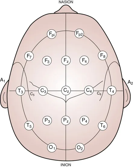
Figure 1-1 10-20 system. A single-plane projection of the head showing all standard positions and the locations of the Rolandic and Sylvian fissures. The outer circle was drawn at the level of the nasion and inion. The inner circle represents the temporal line of electrodes.
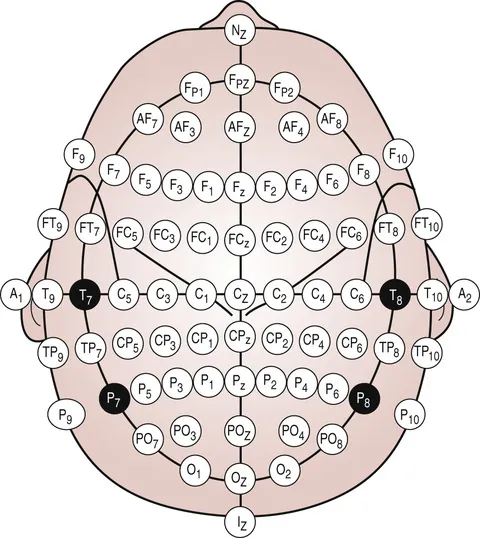
Figure 1-2 10-10 system. The 10-20 system has been modified to standardize a method for adding more electrodes.
Both the 10-10 and the 10-20 system depend on accurate measurements of the skull, utilizing several distinctive landmarks. Essentially, a measurement of the skull is taken in three planes – sagittal, coronal, and horizontal. The summation of all the electrodes in any given plane will equal 100%. Electrodes designat...
Table of contents
- Cover image
- Title page
- Table of Contents
- Copyright
- Foreword
- Dedication
- Preface to the second edition
- 1 Origin and technical aspects of the EEG
- 2 The normal adult EEG
- 3 The normal EEG from neonates to adolescents
- 4 The abnormal EEG
- 5 The EEG and epilepsy
- 6 The EEG in other neurological and medical conditions and in status epilepticus
- 7 The EEG: Tips on indications, reading, and reporting
- Appendix 1 Influence of common drugs on the EEG and on seizure threshold
- Appendix 2 Treatment of Status Epilepticus
- Glossary
- Index
- Questions
- Answers
Frequently asked questions
Yes, you can cancel anytime from the Subscription tab in your account settings on the Perlego website. Your subscription will stay active until the end of your current billing period. Learn how to cancel your subscription
No, books cannot be downloaded as external files, such as PDFs, for use outside of Perlego. However, you can download books within the Perlego app for offline reading on mobile or tablet. Learn how to download books offline
We are an online textbook subscription service, where you can get access to an entire online library for less than the price of a single book per month. With over 1.5 million books across 990+ topics, we’ve got you covered! Learn about our mission
Look out for the read-aloud symbol on your next book to see if you can listen to it. The read-aloud tool reads text aloud for you, highlighting the text as it is being read. You can pause it, speed it up and slow it down. Learn more about Read Aloud
Yes! You can use the Perlego app on both iOS and Android devices to read anytime, anywhere — even offline. Perfect for commutes or when you’re on the go.
Please note we cannot support devices running on iOS 13 and Android 7 or earlier. Learn more about using the app
Please note we cannot support devices running on iOS 13 and Android 7 or earlier. Learn more about using the app
Yes, you can access Rowan's Primer of EEG E-Book by Lara V. Marcuse,Madeline C. Fields,Jiyeoun Jenna Yoo,Ji Yeoun Jenna Yoo in PDF and/or ePUB format, as well as other popular books in Medicina & Neurología. We have over 1.5 million books available in our catalogue for you to explore.