Poultry are a major source of valuable high-quality protein for much of the world's population, so food security is heavily dependent on maintaining poultry health. They are also increasingly important as specialist hobby animals in back-yard flocks. Despite this, veterinarians specializing in the care and health of these important domestic animals are few and far between, and many vets in small animal practice have little real experience of poultry health management and disease. Providing a comprehensive overview, this new handbook will help to plug this gap with 46 chapters of practical and accessible poultry health and management. The book: Covers the poultry industry, basic avian biology, infectious and non-infectious diseases and their agents, infection control, and disease investigation and legislation. Includes full colour images for ease of identification and diagnosis, in addition to practical guides to disease prevention. Considers areas of increasing global importance, such as antimicrobial resistance. Written by international experts, this book forms a valuable illustrated resource for veterinary professionals, veterinary students, or those entering the poultry industry.
eBook - ePub
Poultry Health
A Guide for Professionals
- 352 pages
- English
- ePUB (mobile friendly)
- Available on iOS & Android
eBook - ePub
Poultry Health
A Guide for Professionals
About this book
Trusted by 375,005 students
Access to over 1.5 million titles for a fair monthly price.
Study more efficiently using our study tools.
Information
Topic
MedicineSubtopic
Veterinary Medicine1 Basic Anatomy and Physiology
DAVID PARSONS*
Poultry Health Centre, Trowbridge, Wiltshire, UK
1.1 Introduction
There are important differences between avian and mammalian anatomy as well as between different avian species. Comparison should be made between the external anatomy of the domestic fowl with that of the domestic turkey, domestic duck and domestic goose. The anatomical differences are matched by adaptations in avian physiology.
Diseases of the alimentary, reproductive and respiratory systems are common. An understanding of the anatomy and physiology of domestic poultry underpins the understanding of the spread of infection within a flock and the pathogenesis of the disease in the individual bird.
The anatomy and physiology of these systems are discussed based on Gallus gallus domesticus, the domestic fowl.
1.2 The Respiratory System
The respiratory tract consists of the upper airways: the nostrils, nasal cavity, sinuses and larynx, and lower structures (trachea, lungs and air sacs) (Fig. 1.1).
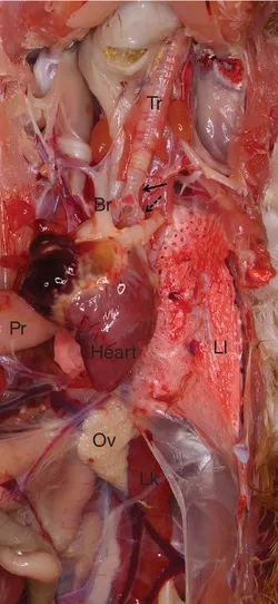
Fig. 1.1. The respiratory tract and other organs visible during initial dissection of the thorax and removal of the rib cage. Tr – trachea; Br – bronchus; L1 – left lung; Pr – proventriculus; Ov – ovary; Lk – left kidney. Small arrow = syrinx. Dotted arrow = primary bronchus. (Author’s own image.)
The nostrils, with the keratinized operculum and cartilaginous lamella, open into the nasal cavity, containing the rostral, middle and caudal conchae. The nasal cavity is separated into two halves by the nasal septum. The choanal cleft is a fissure in the midline of the palate which connects the nasal cavity with the oropharynx. The infraorbital sinus lies laterally and rostroventrally to the eye in the upper jaw. It connects with the nasal cavity ventral to the caudal concha. The nasolacrimal duct opens rostral to the choana and ventral to the middle concha and drains lacrimal secretions from the eye. As in mammals the nasal cavity has important olfaction, air filtration and thermoregulation functions.
Air passes through the choana to the larynx or through the oropharynx in open mouthed breathing to the larynx. The glottis closes reflexly when swallowing to prevent feed entering the trachea. The trachea is made up of about 120 complete signet ring shaped interlocking cartilages that link the larynx to the syrinx which is the voice box and junction with the primary bronchi that connect to the lungs. Branches from the primary bronchi give rise to smaller diameter secondary bronchi, then parabronchi which connects with the atria. The atria are small diverticula that lead to the air capillaries which are the site for gas exchange.
The paired, wedge shaped lungs lie dorsal to the heart. There is no diaphragm. Consequently, the air in the airways of the thoraco-abdomen approximates atmospheric pressure. The lungs are small and non-distensible. The lung can be divided into two parts called the paleopulmo and the neopulmo. In the former, the airflow is unidirectional whilst in the latter it can flow in either direction according to the stage of the respiratory cycle.
The primary bronchus extends to the caudal edge of the lung linking to the ostia from which the abdominal air sac arises. There are nine air sacs in the fowl: a single clavicular and paired cervical, cranial and caudal thoracic and abdominal air sacs (Table 1.1). Their function is ventilation and reducing body weight for flight. The sacs are lined by a single epithelial layer surrounded by connective tissue and are very poorly vascularized. The air sacs arise from secondary bronchi. The exception is the abdominal air sac which has connections with the primary and secondary bronchi.
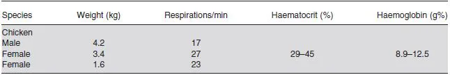
Table 1.1. Air sac volumes in domestic fowl. (Adapted from Sturkie, 1986.)
| Air sacs | Volume air (ml) (2.9 kg chicken) |
|---|---|
| Cervical × 1 | 20 |
| Clavicular × 1 | 55 |
| Cranial thoracic × 2 | 50 |
| Caudal thoracic × 2 | v24 |
| Abdominal × 2 | 110 |
| Lungs × 2 | 35 |
| Skeletal | 4 |
| Total | 298 |
The cervical air sac lies between the lungs and dorsal to the oesophagus. Diverticula extend into the cervical vertebrae along the neural canal and outside to the level of the axis. The clavicular air sac comprises both intra- (around the heart and along the sternum) and extra-thoracic (between bones and muscles of the thoracic girdle and shoulder joints) diverticula. Oesophagus, syrinx, trachea, associated muscles, nerves and blood vessels are suspended in the clavicular or between the clavicular and cervical air sacs. The cranial and caudal thoracic air sacs have no diverticula. The abdominal air sacs have perirenal diverticula to the kidney, pelvis, synsacrum, free thoracic vertebrae and femoral diverticula to bones and muscles of the pelvic limb.
The poor vascularization and absence of cilia make removal of purulent material and treatment of this area difficult. This also facilitates spread of infection throughout the body via the diverticula or directly across the membrane. Thus, airsacculitis detected at slaughter means rejection of the whole carcass.
The cranial group of air sacs (cervical, clavicular and cranial thoracic) contains almost as much air as the caudal group (caudal thoracic and abdominal). Most respiratory movement occurs caudally. Therefore, on inspiration most air enters the caudal air sacs.
In the domestic fowl, the following are pneumatized: subcutaneous facial planes and between skeletal muscle, humerus, coracoid, sternum, ribs, synsacrum, pelvis, cervical and thoracic vertebrae.
Circulation of air through the respiratory tract requires two breathing cycles. Inspiration draws fresh air into the posterior air sacs (caudal thoracic and abdominal air sacs) and oxygen depleted air into the anterior air sacs (the cervical, clavicular and cranial thoracic air sacs). Expiration pushes fresh air from caudal thoracic and abdominal air sacs into the lungs and oxygen depleted air from the cervical, clavicular and cranial thoracic air sacs into the bronchi and trachea to be breathed out.
It is important to remember that holding a bird upside down or holding a bird too tightly will cause respiratory distress and possibly death.
Breathing (Table 1.2) is controlled by the central nervous system (CNS) with sensory inputs from (i) central chemoreceptors – identified in mammals and probably present in birds; (ii) arterial chemoreceptors; (iii) intrapulmonary chemoreceptors and others including: (iv) air sac mechanoreceptors; (v) thermal receptors in the spinal cord; (vi) upper airway receptors sensitive to irritants, cold and possibly (vii) arterial baroreceptors.
Table 1.2. Respiratory parameters in domestic fowl. (Adapted from Sturkie, 1986.)
Increasing ambient CO2 will increase ventilation. However, ventilation is also influenced by exercise, hot or cold temperatures and altitude. There is a balance between the stimulatory and inhibitory drive to ventilate. Hence, in hot weather gular fluttering is important because heat is lost with minimum active ventilation.
1.3 The Alimentary System
In addition to its role in nutrition, the alimentary system is also the largest immunological organ (see Chapter 2 this volume) as well as being host to a vast microbiome (see Chapter 17 this volume). The alimentary tract is short and antiperistalsis is an important compensation in digestion. Many growth and developmental changes occur soon after the chick hatches. Managing the development of the alimentary tract to maturity is vital in promoting and maintaining good bird health and welfare. (See Fig. 1.2 for general structure.)
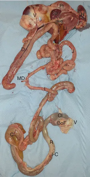
Fig. 1.2. The alimentary tact exposed. Pr – proventriculus; V – ventriculus (gizzard); Sp – spleen; Li – liver; H – heart; D – duodenum; J – jejunum; MD – Meckel’s diverticulum; I – ileum; C – caeca; I-C – ileo-caecal junction; R – rectum; Cl – cloaca; BoF – Bursa of Fabricius; V – vent. The crop is not shown but is located immediately caudal to the proventriculus. (Author’s own image.)
The beak consists of a hard, keratinized epidermal covering which, at its edges, is continually worn away and replaced, and is supplied with extensive sensory innervation, particularly at the tip. The egg tooth is a pointed protuberance on the upper beak which drops off after hatching.
The mouth and pharynx are known as oropharynx. There is no soft palate or pharyngeal isthmus. The choana in the roof of the mouth links oral and nasal cavities. The infundibular cleft behind this is the common opening of the auditory tubes. The salivary glands are located in the roof, cheeks and floor of the mouth. The fowl has about 300 taste buds on the epithelium of the upper beak, and the base and ventrolateral surface on the tongue, with broiler breeds having approximately twice as many as layer breeds.
Food passes along the oesophagus to the crop which is thin walled and distensible. It is a storage organ where bacterial fermentation takes place, reducing the pH to about 4–5.
The proventriculus immediately cranial to the gizzard, produces hydrochloric acid and pepsinogen which is converted to pepsin in the low pH (pH 1–4 depending on food content).
The gizzard (ventriculus) has thick and muscular walls, with a thick yellow to green cuticle on the mucosal surface to protect it from acid and mechanical damage. Finely ground feed – low in fibre – results in poor muscular development while whole grain and high fibre promote good muscular development. Insoluble grit will lodge in the gizzard to facilitate grinding.
The U-shaped duodenum is the first section of the small intestine, enclosing the pancreas in the loop and with a pH of 5.7–7.2. The remains of the yolk sac presents as a small stub, Meckel’s diverticulum, which is used as a marker to separate the jejunum from the ileum. Digestion and absorption of carbohydrates, fats, proteins and also water absorption take place in the small intestine.
The large, paired caeca are attached to the intestine via a sphincter at the ileo-rectal junction. A small enlargement in the neck of the caecum is the caecal tonsil which is a lymphoid organ sampling the microorganisms that enter and leave the caeca. The caeca are filled by the convergence of peristaltic contractions from the small intestine and antiperistaltic contractions from the cloaca and rectum. Liquid only enters the caeca, and the pH varies from 5.7 to 9. The caeca evacuate between one and twelve times daily. Its function is unclear since birds thrive after its removal but some hind-gut fermentation and water reabsorption are thought to take place.
The short rectum has a role in water conservation and carries urine to the caeca by antiperistalsis.
The cloaca is relatively complex for its size with the coprodeum separated from the urodeum which is the smallest chamber into which the urinary and reproductive tracts empty. This is separated by the uroproctodeal fold from the proctodeum which has an external opening through the vent. In the juvenile there is an opening dorsally to the Bursa of Fabricus, the important immunological organ where B cells mature (see Chapter 2 this volume).
Two important additional organs involved in nutrition are the liver and pancreas. The liver is a key organ manufacturing carbohydrates, proteins and fats and is important in detoxification. It also generates heat, r...
Table of contents
- Cover
- Half Title
- Title
- Copyright
- Contents
- List of Contributors
- Preface
- Section 1: Poultry and the Poultry Industry
- Section 2: Infectious Diseases of Poultry
- Section 3: Control of Poultry Diseases
- Index
- Cabi
- Back
Frequently asked questions
Yes, you can cancel anytime from the Subscription tab in your account settings on the Perlego website. Your subscription will stay active until the end of your current billing period. Learn how to cancel your subscription
No, books cannot be downloaded as external files, such as PDFs, for use outside of Perlego. However, you can download books within the Perlego app for offline reading on mobile or tablet. Learn how to download books offline
Perlego offers two plans: Essential and Complete
- Essential is ideal for learners and professionals who enjoy exploring a wide range of subjects. Access the Essential Library with 800,000+ trusted titles and best-sellers across business, personal growth, and the humanities. Includes unlimited reading time and Standard Read Aloud voice.
- Complete: Perfect for advanced learners and researchers needing full, unrestricted access. Unlock 1.5M+ books across hundreds of subjects, including academic and specialized titles. The Complete Plan also includes advanced features like Premium Read Aloud and Research Assistant.
We are an online textbook subscription service, where you can get access to an entire online library for less than the price of a single book per month. With over 1.5 million books across 990+ topics, we’ve got you covered! Learn about our mission
Look out for the read-aloud symbol on your next book to see if you can listen to it. The read-aloud tool reads text aloud for you, highlighting the text as it is being read. You can pause it, speed it up and slow it down. Learn more about Read Aloud
Yes! You can use the Perlego app on both iOS and Android devices to read anytime, anywhere — even offline. Perfect for commutes or when you’re on the go.
Please note we cannot support devices running on iOS 13 and Android 7 or earlier. Learn more about using the app
Please note we cannot support devices running on iOS 13 and Android 7 or earlier. Learn more about using the app
Yes, you can access Poultry Health by Paul Barrow, Venugopal Nair, Susan Baigent, Robert Atterbury, Michael Clark, Paul Barrow,Venugopal Nair,Susan Baigent,Robert Atterbury,Michael Clark in PDF and/or ePUB format, as well as other popular books in Medicine & Veterinary Medicine. We have over 1.5 million books available in our catalogue for you to explore.