Diatom Morphogenesis
eBook - ePub

Diatom Morphogenesis

Vadim V. Annenkov, Joseph Seckbach, Richard Gordon, Vadim V. Annenkov, Joseph Seckbach, Richard Gordon

Share book
  1. English
  2. ePUB (mobile friendly)
  3. Available on iOS & Android
eBook - ePub

Diatom Morphogenesis

Vadim V. Annenkov, Joseph Seckbach, Richard Gordon, Vadim V. Annenkov, Joseph Seckbach, Richard Gordon

Book details
Book preview
Table of contents
Citations

About This Book

DIATOM MORPHOGENESIS

A unique book presenting the range of silica structures formed by diatoms, theories and hypotheses of how they are made, and applications to nanotechnology by use or imitation of diatom morphogenesis.
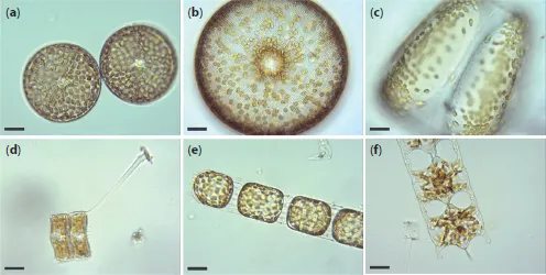
There are up to 200, 000 species of diatoms, each species of these algal cells bearing an ornate, amorphous silica glass shell. The silica is structured at 7 orders of magnitude size range and is thus the most complex multiscalar solid structure known. Recent research is beginning to unravel how a single cell marshals chemical, physical, biochemical, genetic, and cytoskeletal processes to produce these single-cell marvels. The field of diatom nanotechnology is advancing as this understanding matures.

Diatoms have been actively studied over the recent 10-20 years with various modern equipment, experimental and computer simulation approaches, including molecular biology, fluorescence-based methods, electron, confocal, and AFM microscopy. This has resulted in a huge amount of information but the key stages of their silica morphogenesis are still not clear. This is the time to reconsider and consolidate the work performed so far and to understand how we can go ahead.

The main objective of this book is to describe the actual situation in the science of diatom morphogenesis, to specify the most important unresolved questions, and to present the corresponding hypotheses. The following areas are discussed:

  • A tutorial chapter, with a glossary for newcomers to the field, who are often from outside of biology, let alone phycology;
  • Diatom Morphogenesis: general issues, including symmetry and size issues;
  • Diatom Morphogenesis: simulation, including analytical and numerical methods for description of the diatom valve shape and pore structure;
  • Diatom Morphogenesis: physiology, biochemistry, and applications, including the relationship between taxonomy and physiology, biosilicification hypotheses, and ideas about applications of diatoms.

Audience

Researchers, scientists, and graduate students in the fields of phycology, general biology, marine sciences, the chemistry of silica, materials science, and ecology.

Frequently asked questions

How do I cancel my subscription?
Simply head over to the account section in settings and click on ā€œCancel Subscriptionā€ - itā€™s as simple as that. After you cancel, your membership will stay active for the remainder of the time youā€™ve paid for. Learn more here.
Can/how do I download books?
At the moment all of our mobile-responsive ePub books are available to download via the app. Most of our PDFs are also available to download and we're working on making the final remaining ones downloadable now. Learn more here.
What is the difference between the pricing plans?
Both plans give you full access to the library and all of Perlegoā€™s features. The only differences are the price and subscription period: With the annual plan youā€™ll save around 30% compared to 12 months on the monthly plan.
What is Perlego?
We are an online textbook subscription service, where you can get access to an entire online library for less than the price of a single book per month. With over 1 million books across 1000+ topics, weā€™ve got you covered! Learn more here.
Do you support text-to-speech?
Look out for the read-aloud symbol on your next book to see if you can listen to it. The read-aloud tool reads text aloud for you, highlighting the text as it is being read. You can pause it, speed it up and slow it down. Learn more here.
Is Diatom Morphogenesis an online PDF/ePUB?
Yes, you can access Diatom Morphogenesis by Vadim V. Annenkov, Joseph Seckbach, Richard Gordon, Vadim V. Annenkov, Joseph Seckbach, Richard Gordon in PDF and/or ePUB format, as well as other popular books in Biological Sciences & Biology. We have over one million books available in our catalogue for you to explore.

Information

Year
2021
ISBN
9781119488194
Subtopic
Biology
Edition
1

Part 1
GENERAL ISSUES

1
Introduction for a Tutorial on Diatom Morphology

Kalina Manoylov1* and Mohamed Ghobara2
1Dept. of Biological & Environmental Sciences, Georgia College and State University, Milledgeville, GA, United States
2Department of Physics, Freie Universitat Berlin, Berlin, Germany
Abstract
Diatoms are an exceptionally successful group of unicellular microalgae with a large contribution of global primary production in aquatic environments and contributing a significant amount of oxygen to both hydro- and atmospheres. They are fascinating throughout their life and even after death, thanks to their unique cell walls made from ornamented silica. The diatoms include centric species, which may have radial or polar symmetry, and pennates, which include araphid, monoraphid, and biraphid species. Several applications have utilized diatomite, i.e., the fossil form of diatom frustules. To date, many diatomsā€™ secrets have been understood; however, there are still more hidden. Thus, there is a need for more research on diatom basic biology and applications. Seeking this goal, more people should be encouraged to work on diatoms. Often novice researchers are overwhelmed by the terminology associated with the diverse morphology, the discrepancy between expected features for published descriptions, and the actual observation of those complex 3D organisms, which can be a barrier for more progress. Here, we provide a brief introduction to the beginners with a guide to approach the complex diatom morphology focusing on the tools that can be used for its study.
Keywords: Diatom morphology, tutorial, LM and SEM, frustule morphology

1.1 Diatoms in Brief

Diatoms are unicellular, eukaryotic, microscopic algae (range from 1.5 Ī¼m to 5 mm in length, or diameter [1.9]), which maintain large population numbers and contribute considerably to the carbon and oxygen cycle on a global scale [1.8]. This ecologically successful group of algae is present in all aquatic habitats e.g. [1.1, 1.2] and even extends to humid terrestrial places. In aquatic habitats, diatoms are present in the photic zone, i.e., the region of water that light strongly penetrates, as well as in the benthic zone, i.e., the lowest level of water adjacent to the bottom with dim light conditions, depending on water column height and waterā€™s turbidity. Diatoms can exist as planktonic (i.e., suspended in the water column), benthic (i.e., living near the bottom), epiphytic (i.e., adhered to aquatic plants [1.19], Figures 1.2cā€“d), or epizoic (i.e., adhered to a wide range of marine organisms such as crustaceans, mollusks, and vertebrates [1.19, 1.38]), or epilithic (i.e., attached completely or partially to submerged rocks). The adhesion ability of some diatoms is related to their mucilage secretion from specialized areas within their rigid cell walls (such as examples shown in Figures 1.1d and 1.2cā€“d). Some diatoms can form colonies in different arrangements such as chains and ribbons (examples shown in Figures 1.1 and 1.2).
Diatoms are a unique group of microalgae for several reasons, but one of the most notable and unique differences is the glass cell walls they possess [1.45]. This cell wall is called the ā€œfrustuleā€ and is composed of amorphous hydrated silica that gives it unique properties. In general, the frustule is composed of two pieces that fit together like a petri-dish, meaning that the lower part of the frustule, called the hypotheca, sits inside of the upper part of the frustule, called the epitheca. The frustule volume extends by adding strips of silica called girdle bands (cingulum) to the mantle, i.e., the curved edge of the valve. It should be noted that there are plenty of frustule morphologies that vary between taxa.
Diatoms reproduce both asexually (visible in Figure 1.6) and sexually. Most of the time, they reproduce asexually via binary fission through adding new hypovalves to the parent valves. Those new hypovalves are synthesized inside the silica deposition vesicle (SDV). Only after the new hypovalves have completely synthesized and the protoplast cleavage, as well as the exocytosis of siliceous parts, has occurred, the final splitting apart will occur, leaving two daughter diatoms in place. Because the SDV forms inside of each new cell before splitting into two, each new cell creates a new interior of the petri-dish structure. What this means is that the cell that originally contained the upper part of the petri dish (the epitheca) remains the same size, whereas the cell that originally contained the lower part of the petri-dish (the hypotheca) becomes smaller, since it has now built a smaller hypovalve to fit into it. Repeated cell division, therefore, leads to some part of the resulting population becoming smaller and smaller. Were asexual reproduction the only method by which diatoms reproduces, this could lead the population eventually to become vulnerable to dying out, but diatoms are ingenious and have gotten around this problem. At some point, sexual reproduction is initiated by a number of steps, including meiotic divisions to produce male and female gametes. These cells can find each other, fuse to form a zygote and create a structure known as an auxospore, out of which a new large cell of the diatom species will form, restoring its optimal size, which also depends on the environmental circumstances surrounding the auxospores. Some new research proposes chemical communication with pheromones between the male and female gametes [1.20].
Photos depict living diatoms as observed under LM, brightfield.
Figure 1.1 Living diatoms as observed under LM, brightfield. (a) Two living ...

Table of contents