Portions of this introduction are reproduced by permission from John Wiley and Sons: Diogo, R., Smith, C.M. and Ziermann, J.M. 2015. Evolutionary developmental pathology and anthropology: A new field linking development, comparative anatomy, human evolution, morphological variations and defects, and medicine. Dev Dyn 244:1357–1374. © 2015 Wiley Periodicals, Inc.
Why Do Humans Have Anatomical Variations and Anomalies?
When Charles Darwin wanted to convince a highly skeptical scientific community and general public that we evolved from other primates in The Descent of Man (1871), he started by discussing human anatomical variations and anomalies. Why? Because he knew that a strong way to show that we descend from other animals is to highlight the commonalities we share with them and particularly, the commonalities that are only present in some humans, as a reminder of the nonhuman ancestors we had several millions of years ago. Our anatomical variations and anomalies, in particular those that are related to the presence of evolutionary reversions, are indeed the most direct, strong evidence of our evolutionary past. This is because there are features that are present in adults of other species that are typically absent in humans, but for some reason can still be found in some human adults. The most likely scientific explanation, Darwin argued, is that such features were present in our adult ancestors and then were evolutionary lost but evolutionarily “re-acquired.” Not only that: even for some anatomical features that appear in humans that have nothing to do with traits typically present in other animals, these usually also show that we share evolutionary and developmental commonalities with them because such variations and anomalies are also present in some cases within other species. For instance, one of the more severe malformations in humans is cyclopia, the presence of a single eye. We never had an ancestor that was truly a cyclops, but many other mammals also have cyclopic malformations.
This phenomenon is now known as Alberch’s (1989) ill-named “logic of monsters.” According to this theory, which has been strongly supported by a plethora of empirical data, there is a parallel between the variation/anomalies in normal/abnormal individuals of a certain taxon and the fixed diversity observed in wildtype individuals of other taxa. According to Alberch (1989), this parallel might be achieved through regulation of a conserved developmental program (i.e., a set of genetic and/or epigenetic interactions) such that the structure of these internal interactions constrains the realm of possible variation upon which selection can operate. This program may lead to the breakdown in the evolution of some clades, but within most clades this would lead to death of the embryos. This leads us to distinguish between variant features and anomalies. By definition, anatomical variants are structures present in the common population, that is, the population that is karyotypically normal and that does not have severe congenital malformations, or so-called “birth defects.” Anatomical anomalies are precisely often the result of genetic mutations or severe congenital malformations, such as cyclopia, a feature that is never found in the common population. However, this does not mean that anatomical anomalies are always more severe than variants. For instance, a certain limb muscle missing in an individual from the common population would be seen as a variant, while the very same feature seen in, let’s say, a person with Down syndrome, would be seen as an anatomical anomaly. According to the “logic of monsters,” this parallel between the variation and anomalies in so-called “normal” vs “abnormal” individuals of a certain taxon is exactly to be often expected.
As noted by Diogo et al. (2015), such a parallel between the more common phenotypic variations seen in the normal human population and malformations seen in birth defects is also to be expected according to Shapiro et al.’s (1983) “lack of homeostasis” model. This model was in large part formulated based on observations of human trisomic individuals and states that the presence of a whole extra functioning chromosome or of a large chromosome segment causes a disruption of evolved genetic balance. Because of the obligatory integration of the entire genotype, this disruption affects the products of the trisomic chromosome and other chromosomes. This results in decreased physiological and developmental buffering against genetic and environmental forces, which leads to decreased developmental and physiological homeostasis where the pathways and processes that will be the most often and seriously affected are those that are more unstable. This instability thus leads to variations in the normal population. An illustrative example predicted by both the “logic of monsters” and “lack of homeostasis” hypotheses is the presentation of palmaris longus. The absence of palmaris longus (see Fig 3.14) is a common human polymorphism. Polymorphisms are different from variations, because although they are also present in the “normal” population as variants are, they are present in more than 2% of the “normal” population, while variations are present in less than 2%. So, within the “normal” population there are variants—present in less than 2% of the population; polymorphisms—present between 2% and 50% of the population; and the common phenotype—present in more than 50% of the population. In the case of the palmaris longus, this absence of muscle is a polymorphism because the muscle is absent in about ~15%–20% of the “normal” human population. Interestingly, the absence of the muscle is often amplified in humans with severe congenital malformations. For example, palmaris longus was reported to be absent in 74% (105) of 141 defective upper limbs reviewed by Smith et al. (2015), further reinforcing that variants and polymorphisms are often amplified in people with congenital malformations.
However, with exception to this similarity between the “logic of monsters” and the “lack of homeostasis” models, they differ fundamentally in their assumptions and predictions. The “lack of homeostasis” model argues that defects are mostly random and disorganized due to a general lack of homeostasis. The “logic of monsters” predicts, instead, that defects are mostly “logical” and “constrained” because developmental constraints are kept intact by internal homeostasis, even when things go “wrong” in both evolution and embryonic development. That is why the “logic of monsters” model predicts—contrary to the “lack of homeostasis” model—that congenital malformations mirror, or amplify, variations, and that both of them further mirror evolutionary events that occurred in other taxa at completely different geological times. This prediction has been supported by studies demonstrating that similar patterns of intra-specific diversity within a taxon (plasticity) and inter-specific diversity among different taxa usually result from similar developmental mechanisms (Hodin 2000). The “logic of monsters” is thus framed in an “internalist” view of evolution and development that contrasts with the more “externalist” view of adaptationists, who maintain that selection by the external environment is the main evolutionary force. For example, frogs and salamanders tend to exhibit loss of digit one and reduction of digit five due to developmental constraints. This pattern is found in frogs living in different environments that are exposed to different external factors (Alberch and Gale 1985). That is, the recurrent loss/reduction of such digits in frogs seems to have much more to do with internal mechanisms than with specific external conditions, although of course the latter might play some role in the specific frog groups and/or geological time in which those loses/reductions happen.
The works of Alberch, as well as of his colleague Stephen Jay Gould and various other authors in the 1970s and 1980s, have led to the rise of evolutionary developmental biology (Evo-Devo), which has revived interest in comparative anatomy, particularly of soft tissues, and of “teratology,” now designated as the study of congenital malformations. These are fields that were somewhat dormant for many years after having a prominent role in anatomical and biological studies in the 18th, 19th, and early 20th centuries, with only a few notable works in the latter half of the 20th century (e.g., Willis 1958). Following this trend, one of us (RD) has created, with other colleagues a new subfield within Evo-Devo: Evolutionary Developmental Pathology (Evo-Devo-Path), which is exactly focused on understanding the evolutionary and developmental basis of human variations and anomalies (Diogo et al. 2015; Diogo and Wood 2016). This new subfield—which was specifically applied to human anatomy, by Diogo et al. (2016)—is in a way a comeback to the study of the links between normal and abnormal development and pathologies, which began to be intensively undertaken centuries ago by authors such as St. Geoffroy St. Hilaire, but that were then somewhat neglected for decades during the 20th century. Fortunately, more and more authors are realizing the importance of investigating these links, although with Evo-Devo they have been mainly studied by focusing principally on osteological or superficial features (e.g., absence of a certain bone, shape of head), with fewer studies being done about the muscular system of both humans and nonhuman animals with anatomical variations or anomalies. However, now that we understand the developmental—including both genetic and epigenetic—and evolutionary basis for why we have anomalies and variations, it is time to develop Evo-Devo-Path to the detailed study of soft tissues such as muscles.
For instance, we now know that evolutionary reversions did play a substantial role in primate/human evolution. In a phylogenetic work about muscle evolution in primates, Diogo and Wood (2012b) have shown that 28 out of the 220 (12.7%) evolutionary changes unambiguously optimized in the most accepted primate phylogenetic tree are reversions to a plesiomorphic (ancestral) state. Of those 28 reversions, six were directly related to our own evolution because they occurred at nodes that lead to the origin of modern humans. Nine of these reversions go against Dollo’s law, which states that once a complex structure is lost, it is unlikely to be regained. Our Evo-Devo-Path studies support the idea that reacquisition of morphological structures in adults that have been absent for long periods of time is possible because the associated developmental pathways were maintained in the members of that taxon.
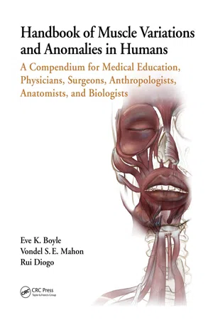
For example, chimpanzees display a reversion of a synapomorphy of the Hominidae (great apes and modern humans) that was acquired at least 15.4 million years ago. Adult chimpanzees have two contrahentes digitorum muscles (see Figure 3.18) in addition to adductor pollicis (see Figure 3.19), which is the only contrahens muscle present in the typical phenotype in adults of other apes. Chimpanzees thus have contrahentes muscles going to digit four and the other to digit five. Developmental studies of hand muscles show that karyotypically normal human embryos do exhibit contrahentes going to various fingers, but these muscles usually are reabsorbed or incorporated within other structures during later embryonic development (Cihak 1972). In karyotypically abnormal humans, such as those with trisomies 13, 18, or 21, the contrahentes often persist after birth as “atavisms” (see Figure 3.18), which are structures that present as developmental anomalies or variations and resemble the common adult character state of the ancestors of the taxon to which the individual belongs. Cihak (1972) showed that intermetacarpales muscles of the hand are also present as discrete muscles in early stages of karyotypically normal human embryonic development but later join with flexor breves profundi muscles to form the dorsal interossei (see Figure 3.18). Therefore, the evolutionary reversions that result in the presence of contrahentes and intermetacarpales muscles in chimpanzees are likely related to heterochronic (specifically paedomorphic) events in the chimpanzee lineage (Diogo and Wood 2012b). So, it is very likely that many variants and anomalies that are present in adult humans and that were present in our adult ancestors are structures that were always present in the embryos of our ancestors, but that then became absent, or fused with other structures, in later stages of development. But, for some reason, either genetic or epigenetic, they persist until later stages of development in certain people as variations or anomalies. That is precisely what happens to features such as the presence of various hand or foot contrahentes muscles, or a platysma cervicale muscle of the head (see Figure 2.3).
We have been studying cadaveric material from fetal, neonatal, and adult humans with trisomies and from mouse models for Down syndrome (e.g., Ts65Dn) to investigate the developmental mechanisms related to the atypical development of muscles. Research on the skeletons of trisomic individuals has supported the idea that for some features there is in fact a developmental delay. For example, some nasal bones develop after the 24th week in cases of trisomy 21, while in karyotypically normal humans the developmental onset is the 10th week (e.g., Keeling et al. 1997). Interestingly, these studies provided examples of different phenotypic patterns often seen in different trisomies, for instance regarding the axial skeleton in fetuses with trisomy 18 vs fetuses with trisomy 21 (Keeling et al. 1997), further supporting the ill-named “logic of monsters.” One hypothesis we aim to test in future works is that the disappearance of certain muscles during early developmental stages of karyotypically normal humans is related to apoptosis, and that the persistence of these atavistic muscles later in ontogeny in individuals with trisomies 13, 18, and 21 is associated with delayed development specifically due to decreased muscle apoptosis. Some authors suggest that humans with DS show an increase of apoptosis in structures such as neurons, granulocytes, and lymphocytes (e.g., Elsayed and Elsayed 2009). If our studies support the hypothesis that these individuals have decreased muscle apoptosis that leads to the presence of additional muscles in later ontogenetic stages, this will imply a more nuanced pattern of apoptosis (i.e., a mosaic scenario where apoptosis is increased in some tissues and decreased in others). Within the numerous Down syndrome cases listed in the tables of Dunlap et al. (1986), there are 12 supernumerary muscles, as opposed to two absences. Therefore, Down syndrome individuals seem to present with more accessory muscles in general, supporting our hypothesis these individuals may have decreased apoptosis in skeletal muscles. Bersu (1980) also suggested that the persistence of some embryonic muscles in later stages of development of trisomic individuals likely has to do with failure of normal cell death or some other process of involution. If there is indeed a contrast in apoptosis between the nervous system (e.g. more apoptosis of nerve cells) and muscular system (i.e. less apoptosis and presence of extra muscles), our hypothesis might also shed light on the etiology of hypotonia (low muscle tone) that is present in individuals with Down syndrome.
Some authors suggest that cases in which complex structures are formed early in “normal” ontogeny but later become lost/indistinct during development (so-called “hidden variation”), may allow organisms to have greater ontogenetic potential early in development. If faced with external perturbations (e.g., climate change, habitat occupied by new species), evolution can use that potential (adaptive plasticity: e.g., West-Eberhard 2003). However, authors such as Gould (1977, 2002) and Alberch (1989) suggested that these cases support instead a “constrained” (internalist) rather than an “adaptationist” (externalist) view of evolution. This is because it is n...