eBook - ePub
A Complete Guide to the Final FRCR 2B
- 444 pages
- English
- ePUB (mobile friendly)
- Available on iOS & Android
eBook - ePub
A Complete Guide to the Final FRCR 2B
About this book
This book aims to help candidates preparing for the Final FRCR 2B examinations held by the Royal College of Radiologists of the UK, and the Joint Final FRCR/FHKCR Part B Examination for the Fellowship of the Royal College of Radiologists and Hong Kong College of Radiologists. This book provides advice on preparation techniques, followed by dozens of practice cases and images relevant to all three sections of the examination: reporting session, rapid reporting session and oral examination/viva voce. The richly-illustrated book contains images of plain X-rays, CT, MR, US and radionuclide scans, making it particularly useful for candidates who have limited access to teaching or film libraries. A selection of both common and uncommon cases is included, giving candidates a realistic idea of the level of preparation and the breadth and depth of knowledge needed for success. Although primarily focused on the FRCR 2B examination, radiology trainees across the world facing viva and reporting sessions and candidates for American Board Examinations will find this a useful and informative book.
Trusted by 375,005 students
Access to over 1.5 million titles for a fair monthly price.
Study more efficiently using our study tools.
Information
Section II
Chapter 1
Reporting session
Case bag 1
CASE 1.1
Age: 72 years | Gender: Male |
Clinical problem: Back pain following a fall | |
Images in exam case: Plain radiograph (4), CT (2) | |
FINDINGS
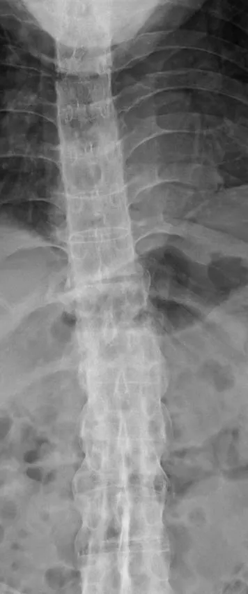
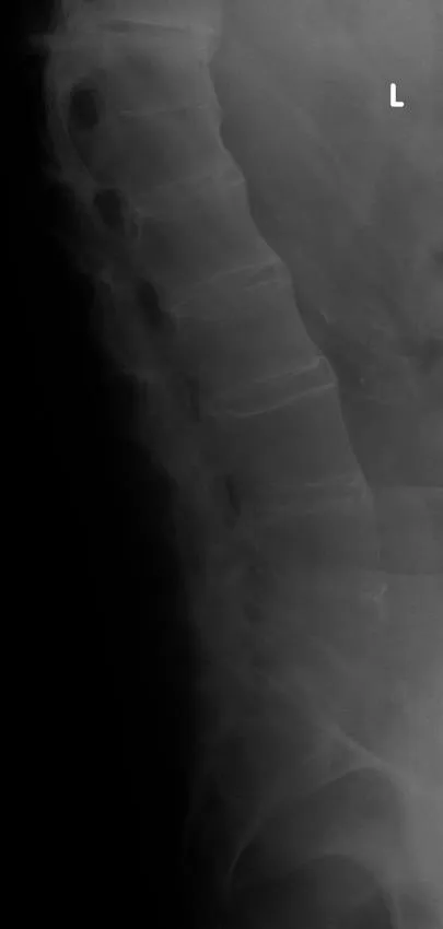
- 1. X-ray thoracolumbar spine:
- a. Marginal syndesmophytes
- b. Ossification of the interspinous ligament – ‘dagger’ sign
- c. Ossification of the anterior longitudinal ligament
- d. Fusion of both sacroiliac joints
- e. Fracture through mid-thoracic intervertebral disc with retrolisthesis of superior vertebra.
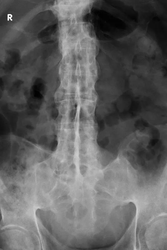
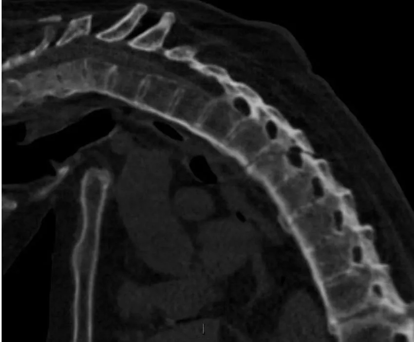
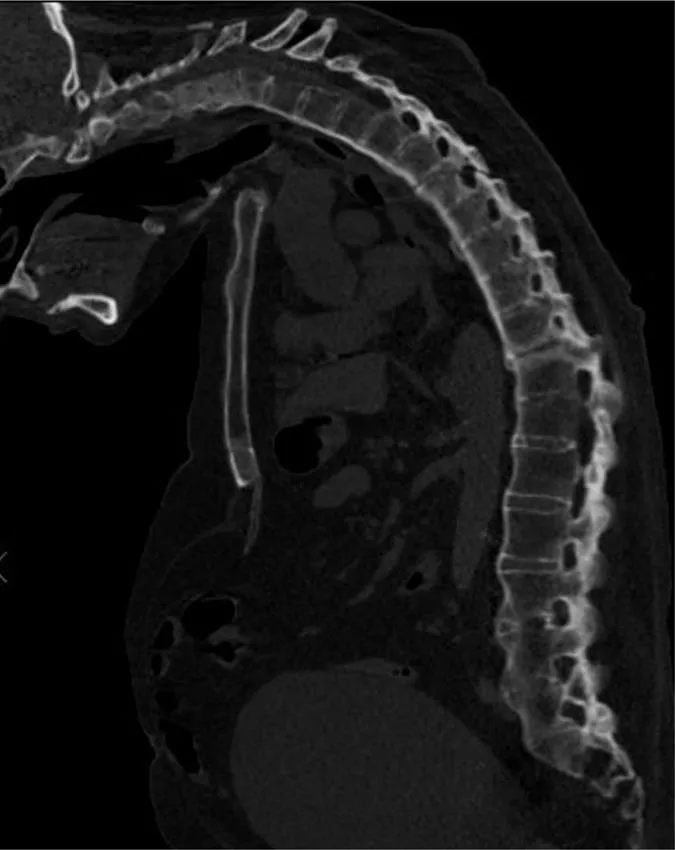
- 2. CT whole spine:
- a. Ossification of anterior longitudinal ligament
- b. Fusion of multiple facet joints
- c. Severe cervicothoracic kyphosis
- d. Fracture line starting at D5/6 intervertebral disc and extending into the posterosuperior body of D6 and D6/7 facet joints
- e. Mild retrolisthesis of D5 over D6
- f. Widened lucent line through D10/11 disc space and facet joints with adjacent reactive sclerosis
- g. Distended urinary bladder.
DIAGNOSIS
- 1. Ankylosing spondylitis, acute fracture through D5/6 extending through the posterior elements. This is an unstable fracture as all three vertebral columns are disrupted.
- 2. Pseudoarthrosis D10/11, most likely secondary to non-union of an old fracture.
FURTHER INVESTIGATIONS AND MANAGEMENT
- 1. Urgent neurosurgical referral, as the fracture is unstable.
- 2. If there are signs of cord compression, MRI may be necessary to look for cord injury or spinal extra-axial haematoma.
FURTHER INFORMATION
Ankylosing spondylitis is a regular feature of the FRCR 2B examination, either as a written case or in the viva. The scenarios involved could be bilateral symmetrical sacroiliac joint erosion, sclerosis or fusion, erosion of the anterosuperior corner of the vertebra on lateral radiograph (Romanus sign), sclerosis of the anterosuperior corner and periostitis of the waist giving rise to vertebral ‘squaring’, syndesmophyte formation leading to ‘bamboo spine’, intervertebral disc calcification, ossification of the anterior longitudinal, posterior longitudinal, interspinous and supraspinous ligaments, kyphosis and facet joint fusion. Other skeletal features include erosions of the symphysis pubis and ischial tuberosities, asymmetrical erosive oligoarthritis, atlanto-axial dislocation and osteoporosis. In a traumatic setting, the fracture line may run through the intervertebral disc space right through into the posterior elements, and may be missed if one is not vigilant. Clinically occult fractures can also occur, leading to mobile non-union (pseudoarthrosis) – the so-called Anderson lesion.
CASE 1.2
Age: 63 years | Gender: Male |
Clinical problem: Histo... | |
Table of contents
- Cover
- Half Title
- Title Page
- Copyright Page
- Table of Contents
- Foreword
- Preface
- List of image contributors
- Dedication
- Section I
- Section II
- Further reading
- Index
Frequently asked questions
Yes, you can cancel anytime from the Subscription tab in your account settings on the Perlego website. Your subscription will stay active until the end of your current billing period. Learn how to cancel your subscription
No, books cannot be downloaded as external files, such as PDFs, for use outside of Perlego. However, you can download books within the Perlego app for offline reading on mobile or tablet. Learn how to download books offline
Perlego offers two plans: Essential and Complete
- Essential is ideal for learners and professionals who enjoy exploring a wide range of subjects. Access the Essential Library with 800,000+ trusted titles and best-sellers across business, personal growth, and the humanities. Includes unlimited reading time and Standard Read Aloud voice.
- Complete: Perfect for advanced learners and researchers needing full, unrestricted access. Unlock 1.5M+ books across hundreds of subjects, including academic and specialized titles. The Complete Plan also includes advanced features like Premium Read Aloud and Research Assistant.
We are an online textbook subscription service, where you can get access to an entire online library for less than the price of a single book per month. With over 1.5 million books across 990+ topics, we’ve got you covered! Learn about our mission
Look out for the read-aloud symbol on your next book to see if you can listen to it. The read-aloud tool reads text aloud for you, highlighting the text as it is being read. You can pause it, speed it up and slow it down. Learn more about Read Aloud
Yes! You can use the Perlego app on both iOS and Android devices to read anytime, anywhere — even offline. Perfect for commutes or when you’re on the go.
Please note we cannot support devices running on iOS 13 and Android 7 or earlier. Learn more about using the app
Please note we cannot support devices running on iOS 13 and Android 7 or earlier. Learn more about using the app
Yes, you can access A Complete Guide to the Final FRCR 2B by Deepak Subedi,Marialena Gregoriades,En Hsun Choi,John T Murchison in PDF and/or ePUB format, as well as other popular books in Medicine & Medical Theory, Practice & Reference. We have over 1.5 million books available in our catalogue for you to explore.