The electrophoresis techniques are used in medicine, biochemistry, analytical chemistry, and biology to separate soluble and insoluble proteins, nucleic acids, chromosomes, viruses, as well as lysosomes, mitochondria, ribosomes and other cell organelles, red cells, tissue cells, and parasites. This book provides a view over the old electrophoresis techniques, as well as the recent developments in electrophoresis.
Electrophoresis Fundamentals is based on the recent book Electrophoresis: Theory and Practice published in 2020 by De Gruyter. The previous book combines theory and technical applications with troubleshooting and problem solving. While Electrophoresis is intended for specialists, Electrophoresis Fundamentals is a book for laboratory technicians, students, biochemists, general practitioners, and more.
Trusted by 375,005 students
Access to over 1.5 million titles for a fair monthly price.
The term electrophoresis means moving of charged dissolved particles in an electric field, which causes their resolution depending on their velocities and interaction with the separation medium [1, 2, 3]. In its current form, the electrophoresis is connected with the studies of Tiselius [4] in the 1930s.
The electrophoresis of positively charged particles (cations) is called cataphoresis; the electrophoresis of negatively charged particles (anions) is called anaphoresis.
The electrophoresis is used for resolving of proteins and nucleic acids, chromosomes, viruses, cell membranes (plasma, lysosomal, nuclear, and other), cell organelles (mitochondria, ribosomes), cells (red cells, tissue cells, and parasites), etc., it takes place in biochemistry, proteomic and genomic studies, forensics, molecular biology, and microbiology. By electrophoresis, more than the half of all separations and almost all separations of blood proteins and DNA are carried out.
Overview on electrophoresis
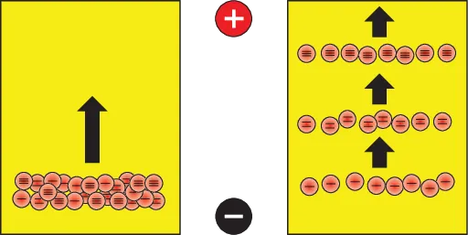
Proteins and nucleic acids form polyions. In an electric field, the positively charged polyions move toward the negative pole, while the negatively charged polyions move toward the positive pole (Figure 1.1).
Left – start of electrophoresis; Right – end of electrophoresis.
Figure 1.1: Electrophoresis of unipolar particles carrying different electric charges.
Commonly, polyions that are to be resolved are applied onto a separation medium, which in its turn is placed into an electrophoresis cell that is connected to a power supply. When electric current is turned on, the larger and less charged polyions move slower through the medium, while the smaller and more charged polyions move faster.
Mostly, electrophoresis is carried out in agarose or polyacrylamide gels, soaked with buffers. Other separation media are: starch gel, cellulose acetate, and paper. Today they have lost their actuality. Electrophoresis can also be carried out in buffers only. The buffers are electrolyte solutions, which maintain constant pH values, e.g. TRIS-borate buffer, TRIS-histidinate buffer, Goods buffers, and more.
Since proteins and nucleic acids are mostly colorless, their movement through the gel cannot be followed during electrophoresis. Therefore, tracking dyes are usually included in the sample buffer. At electrophoresis of negatively charged proteins, Bromophenol blue, Xylene cyanol, which runs slower than Bromophenol blue, or Orange G are used; at electrophoresis of positively charged proteins Bromocresol green or Methylene blue are used.
Proteins can be separated also in pH gradients, where they stop at their isoelectric (pI) points. This electrophoresis, called isoelectric focusing, can be carried out in two types of pH gradients: produced by carrier ampholytes, or by immobilines.
The electrophoresis takes place in horizontal or vertical separation cells. The electrodes can be placed on the gel or in separate electrode tanks.
Beside electrophoresis cells and power supplies, thermostats for reserving the resolving medium temperature, and densitometers or scanners for analyzing the pherograms (the resolved polyions in the medium) are used. Semi-automated electrophoresis devices are also offered on the market.
The electrophoretic velocity of a polyion is proportional to its effective mobility and the electric field strength . In its turn, the effective mobility depends on the total electric charge of the polyion and is inversely proportional to the viscosity of the separation medium. The total electric charge is determined by the buffer pH value; and the viscosity of the resolving medium depends mainly on the medium structure and temperature.
The electric field strength is equal to the ratio between the electric voltage and distance between the two electrodes. Since the distance remains constant during the electrophoresis, the polyion velocity depends only on the electric voltage.
Electrophoresis should be carried out at voltage and electric current, when the heating could be drawn out from the electrophoresis cell.
After electrophoresis, blotting of proteins and nucleic acids can be made. Using this technique, the resolved bands can be immobilized onto blot membranes and treated afterward. The blotting methods are carried out in four steps: electrophoretic separation of proteins or nucleic acid fragments; their transfer and immobilization onto blot membranes; binding of analytical probes to the blotted substances; and visualization of the blotted bands.
The blotting of DNA bands is called Southern blotting, the blotting of RNA bands is called Northern blotting, and the blotting of protein bands is called Western blotting. The blotting membrane consists usually of nitrocellulose, nylon or polyvinylidene difluoride (PVDF).
Separation media
The electrophoresis can be carried out in a solution, but the diffusion there is too strong. In order to limit the diffusion, solid media are used. The earliest solid medium was cellulose contained in the filter paper. The paper electrophoresis was invented by Kunkel and Tiselius [5] in 1951. Cellulose acetate membranes were the next step in the electrophoresis progress. Today most common are agarose and polyacrylamide gel electrophoresis.
Electrophoresis resolution and sharpness
The resolution of electrophoresis is referred to the ability of electrophoresis to separate two sample components from each other. It depends on the Gaussian profiles of the bands and is calculated by dividing the distance between the centers of adjacent bands by their average bandwidths.
The sharpness of electrophoresis is referred to as the reciprocal value of the bandwidth; narrow the bands, the higher the sharpness.
Detection of resolved bands
The majority of polyion bands are not visible to the naked eye, with a few exceptions. Direct optical detection of resolved bands can be applied, for example, to hemoglobins (which are red colored). Therefore, a couple of methods are created for detection, localization, and quantitation of separated bands.
The detection of resolved polyions is carried out directly or indirectly. The direct detection is performed in the resolving medium by nonspecific or specific staining, enzyme-substrate reactions, immune precipitation, autoradiography, and fluorography. The indirect detection is performed by immune printing or blotting.
The proteins can be stained in gels. Common dye is Coomassie brilliant blue, which can detect 0.3 μg of protein in a spot. DNA is detected usually by fluorescent intercalating of ethidium bromide. Both proteins and DNA can react with silver ions to form black bands. The silver methods are 100 times more sensitive than the other staining methods and can detect 2 ng of protein in a spot.
The process of staining is followed by destaining of the gel background to remove unbound dye. In some cases, placing the gel on an ultraviolet (UV) lightbox or under a UV lamp can reveal UV absorbing bands, or UV fluorescence bands.
If radioactive atoms have been incorporated in the polyions prior to electrophoresis, their position in the gel or membrane can be determined by autoradiography. For this purpose, the gel or membrane is placed on a photographic film or overlaid with it and let to expose in the dark and cold to show the positions of the radioactive bands.
The most modern methods of both detection and characterization of resolved proteins involve mass spectrometry.
The electrophoresis results can be recorded with a computer operated camera, and the intensity of a band or spot of interest can be compared against markers in the same gel, using specialized software.
After electrophoresis, the gels with bands can be saved in dry forms.
References
[1]Lyklema J. Fund Interface Colloid Sci, 1995, 2, 3, 208. →
[2]Hunter RJ. Foundations of Colloid Science, 2nd edn, Oxford University Press, Oxford, 2001. →
[3]Russel WB, Saville DA, Schowalter WR. Colloidal Dispersions. Cambridge University Press, Cambridge, 1989. →
[4]Tiselius A. Trans Faraday Soc, 1937, 33, 524–531. →
[5]Kunkel HG, Tiselius A. Electrophoresis of proteins on filter paper, J Gen Physiol, 1951, 35, 89–118. →
Table of contents
Title Page
Copyright
Contents
Preface
About the Author
Abbreviations
1 Fundamentals of electrophoresis
1.1 Electric double layer of a charged particle
1.2 Proteins and nucleic acids form polyions in solution
1.3 Electrophoresis is running in buffers
1.4 Polyions are moving in electric field
1.5 Electrophoresis is carried out in different solid media
1.6 General theory of electrophoresis
1.7 Electrophoresis instrumentation
1.8 Classification of electrophoretic methods
2 Electrophoresis of proteins
2.1 Cellulose acetate electrophoresis of proteins
2.2 Agarose gel electrophoresis of proteins
2.3 Immunoelectrophoresis
2.4 Affinity electrophoresis
2.5 Polyacrylamide gel zone electrophoresis of proteins
2.6 Isotachophoresis of proteins
2.7 Disc-electrophoresis of proteins
2.8 Isoelectric focusing of proteins
2.9 Free-flow electrophoresis of proteins
2.10 Capillary electrophoresis of proteins
2.11 Two-dimensional electrophoresis
2.12 Preparative electrophoresis of proteins
2.13 Microchip electrophoresis of proteins
2.14 Blotting of proteins
2.15 Evaluation of protein pherograms
2.16 Precast gels for protein electrophoresis. Rehydratable gels
3 Electrophoresis of nucleic acids
3.1 Agarose gel electrophoresis of nucleic acids
3.2 Pulsed-field gel electrophoresis of nucleic acids
3.3 Capillary electrophoresis of nucleic acids
3.4 Polyacrylamide gel electrophoresis of nucleic acids
3.5 Microchip electrophoresis of nucleic acids
3.6 Blotting of nucleic acids
3.7 Evaluation of nucleic acid pherograms
3.8 Precast gels for nucleic acid electrophoresis
4 Iontophoresis
5 History of electrophoresis and iontophoresis
6 Troubleshooting
Problems
Solution of problems
Reagents for electrophoresis
Recipes for electrophoresis solutions
SI units and physical constants used in electrophoresis
Electrophoresis terms
Index
Frequently asked questions
Yes, you can cancel anytime from the Subscription tab in your account settings on the Perlego website. Your subscription will stay active until the end of your current billing period. Learn how to cancel your subscription
No, books cannot be downloaded as external files, such as PDFs, for use outside of Perlego. However, you can download books within the Perlego app for offline reading on mobile or tablet. Learn how to download books offline
Perlego offers two plans: Essential and Complete
Essential is ideal for learners and professionals who enjoy exploring a wide range of subjects. Access the Essential Library with 800,000+ trusted titles and best-sellers across business, personal growth, and the humanities. Includes unlimited reading time and Standard Read Aloud voice.
Complete: Perfect for advanced learners and researchers needing full, unrestricted access. Unlock 1.5M+ books across hundreds of subjects, including academic and specialized titles. The Complete Plan also includes advanced features like Premium Read Aloud and Research Assistant.
Both plans are available with monthly, semester, or annual billing cycles.
We are an online textbook subscription service, where you can get access to an entire online library for less than the price of a single book per month. With over 1.5 million books across 990+ topics, we’ve got you covered! Learn about our mission
Look out for the read-aloud symbol on your next book to see if you can listen to it. The read-aloud tool reads text aloud for you, highlighting the text as it is being read. You can pause it, speed it up and slow it down. Learn more about Read Aloud
Yes! You can use the Perlego app on both iOS and Android devices to read anytime, anywhere — even offline. Perfect for commutes or when you’re on the go. Please note we cannot support devices running on iOS 13 and Android 7 or earlier. Learn more about using the app
Yes, you can access Electrophoresis Fundamentals by Budin Michov in PDF and/or ePUB format, as well as other popular books in Biological Sciences & Biochemistry. We have over 1.5 million books available in our catalogue for you to explore.