The Evolution of Radionanotargeting towards Clinical Precision Oncology is a remarkable book honoring Professor Kalevi Kairemo, who is known among academic and medical circles as a pioneer in novel radiolabeled therapeutics. This festschrift provides an overview of key advances in the field of radionanotargeting, and the directions for future development in patient care. Prof Kairemo's research is based on multiomics, which involves multiple elements: genomics, transcriptomics, proteomics, metabolomics, microbiomics, epigenomics, exposome, imaging, and precision medicine, which is reflected by the unique collection of articles presented. The articles start from the angle of radionanotargeting and theragnostics leading to imaging and therapy, which includes sections for thyroid cancer, head and neck cancer, genitourinary cancers and neuroendocrine neoplasms. Theragnostics, nanoparticles and precision oncology have also been covered in their own segments, while also giving a glimpse of research in metabolic imaging, cardiovascular radionuclide imaging, and bone therapies. The sequence of chapters demonstrates how, through Professor Kairemo's efforts, radionanotargeting has evolved to a practice changing therapeutic approach in the clinic, particularly in oncology. Finally, Professor Kairemo's own memoir, "Seven decades in health care" and memoirs from colleagues including a personal introduction to him with a photographic cavalcade reveals the world of a multitasking person with a multidisciplinary approach to science, that ushered his development of significant expertise across the fields of chemistry, biology, engineering, physics and clinical medicine. This book is excellent for medical historians, trainees and specialists in clinical and radiological oncology in expanding their understanding of the role of radionuclide imaging over the years, making it an ideal tribute that belongs in the library of anyone involved in the field.
eBook - ePub
The Evolution of Radionanotargeting towards Clinical Precision Oncology: A Festschrift in Honor of Kalevi Kairemo
- English
- ePUB (mobile friendly)
- Available on iOS & Android
eBook - ePub
The Evolution of Radionanotargeting towards Clinical Precision Oncology: A Festschrift in Honor of Kalevi Kairemo
About this book
Trusted by 375,005 students
Access to over 1.5 million titles for a fair monthly price.
Study more efficiently using our study tools.
Information
“Fit for Purpose” 64Cu Labeled Liposome Formulations Specialized for Enriched Targeting to 1) Bone Marrow Spleen; 2) lymph Nodes; or 3) Tumor
Sang-Gyu Lee1, *, Kishore Pillarsetty1, Steven M. Larson1
1 Larson Lab, Molecular Pharmacology Program, Sloan Kettering Institutes, Memorial Sloan Kettering Cancer Center, New York, NY, USA
Abstract
Upon intravenous injection, liposomal particles are rapidly cleared from the blood by fixed macrophages in the liver, spleen, bone marrow and lymph nodes i.e. specialized cells of the innate immune system. By reducing the affinity for these fixed macrophages, therapeutic liposomes containing drugs have been used to target tumor by the Enhanced Permeability and Retention (EPR) effect. In this communication, we report minor modifications of lipid compositions that result in the production of 3 classes of liposomes, each suitable for enhanced selectivity of uptake in 1) Bone Marrow/Spleen (SBMT Lipo); 2) Lymph Nodes (LNT Lipo); and 3) Tumor (TT Lipo).
Keywords: Bone Marrow Imaging, Copper-64, Endothelial Macrophages, Lymph Node Imaging, Nanoparticles, PET, Reticuloendothelial System, Tumor Imaging.
* Corresponding author Sang-Gyu Lee: Larson Lab, Molecular Pharmacology Program, Sloan Kettering Institutes, Memorial Sloan Kettering Cancer Center, New York, NY, USA; E-mail: [email protected]
Introduction
For many years nuclear medicine has exploited the cells of the fixed macrophage system for imaging purposes by using microscopic particles as radiaotracers. Initially, Au-198 colloid and succeeded by Tc-99m colloid were used to imaging liver, for detection of metastatic tumors, as well as bone marrow to identify sites for biopsy. More recently, a variety of simple radiolabeled particles are used for lymphatic imaging upon subcutaneous injection, i.e. sentinel lymph node imaging for breast cancer and melanoma staging. These have also included a variety of nanoparticles of various materials and sizes.
In the current manuscript, by modulating the size and surface lipid composition,
we have developed liposomal formulations that allow for enhanced targeting to organ-specific cells of the fixed macrophage system (aka Reticuloendothelial System (RES), after parenteral administration. Liposomes in the size range of ~90 nanometers in diameter with varying degrees of pegylation and surface charge were developed for specific targeting purposes. For example, SBMT Lipo preferentially accumulates in the phagocytic cells lining the bone marrow and spleen sinusoids [1]. SBMT Lipo formulation was used to provide enhanced delivery of radioprotector drug γ-tocotrienol (GT3), or GT3-Nano for short, to the spleen and bone marrow, to mitigate bone marrow radiation damage from external beam radiation or targeted radionuclide therapy (TRT). Fig. (1) shows the biodistribution profile of different “fit for purpose” liposomal formulations that can be used for distinct purposes, including imaging, drug delivery.
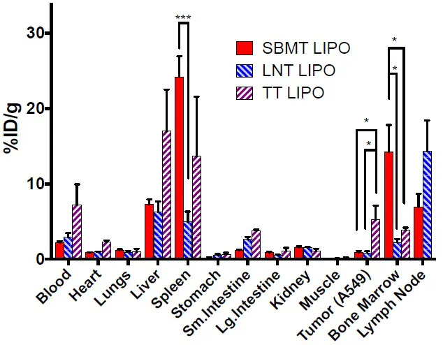
Biodistribution of 64Cu labeled selective targeting liposomes at 24 h post-injection. Athymic nude mice bearing A549 tumor were injected with ~140 μCi (5.18 MBq) of 64Cu labeled liposomes. 1.4 mg (2 μmoles) of lipid was injected each mouse through tail vein injection and it is corresponding to approximately 6 x 10¹² liposome particles per mouse. Mice were euthanized at 24 h post-injection and organs were harvested in pre-weighed tubes to measure organ weight and γ-counting. Data were presented as mean and SEM. n=5 per group. * p < 0.05, *** p < 0.001.
Methods
The compositions of each liposome are as follows; SBMT Lipo. Phosphatidylcholine (PC), cholesterol, succinyl phosphoethanolamine (SuPE) and mPEG2k-DSPE (6:3:1:0.1). LNT Lipo. The ratio was adjusted to 6:3:1:0.7. TT Lipo, the ratio between PC, cholesterol, and mPEG2k-DSPE are 7:3:0.5. Tumor targeting no SPE MPEG 5%. All liposomes were manufactured by the standard extrusion method at 65 oC and the sizes of liposomes are 90 nm. The formulation was stable for 2 years at 4 oC in size and 64Cu labeling to the liposomes. Labeling was performed with 64Cu chelation to DOTA-bn- DSPE doped liposomes after incubation of [64Cu] CuCl2 at 37 C for 1 hour. ITLC was used for quality control, and yield was quantitative [2].
Animal studies. Mice were injected intravenously by tail vein and ex-vivo biodistribution was performed at 24 hours post-injection. Mice were imaged 24 h post liposome administration using Focus 120 microPET or Inveon PET/CT scanner.
Results and Discussion
Using 3 different formulations, mice were sacrificed, and biodistribution was performed at 24 hours after injection of approximately 6x10¹² particles (60 pmoles), of liposomal preparations of approximated 90 nm in diameter of 3 stable formulation types: SBMT Lipo, LNT Lipo, and TT Lipo. 64Cu, a positron emitter, provides a convenient radiotracer for gamma counting and/or PET imaging. (1) A comparison of the biodistribution results from independent studies is shown in Fig. (1). All 3 formulations target the spleen, bone marrow, liver and lymph nodes. However, by altering the surface charge and/or pegylation fraction, major changes in relative biodistribution occur. As is clear from Fig. (1), there are statistically significant distribution differences between the tissues, and this translates into major differences in PET imaging appearance. In Fig. (1), in one case, a tumor was targeted and comparison, in this case, is between the SBMT Lipo and the TT Lipo. Tumor uptake is most favorable in this TT Lipo formulation, with a mean change greater than 5 times the uptake in tumors seen with the SBMT LIPO.
The LNT Lipo steers liposomes to the lymph node, as shown in Fig. (2A). The SBMT Lipo formulation steers the liposome to the spleen and bone marrow at the expense of the liver uptake Fig. (2B). The preparation was developed to deliver cargo as selectively as possible to the spleen and bone marrow, wherein stem cells reside that are important for the regeneration of bone marrow, damaged by radiation effects. A recent publication in the JNMMI [2] describes a newly developed radioprotector drug liposomal formulation GT3-Nano based on SBM Lipo formulation that carriers radioprotector Gamma-tocotrienol (GT3), a form of Vitamin E,. This formulation promotes recovery of hematopoietic function post-radiation. Among the uses will be protected against radiation toxicity from TRT approaches under active development in multiple clinical trials. This protection may afford higher dose delivery to achieve greater efficacy of TRT in man While minimizing toxic effects on bone marrow.
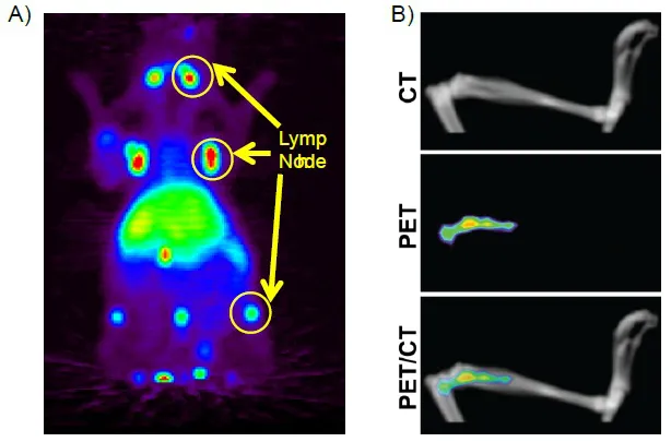
MIP images of mice labeled with 64Cu to A) lymph node targeting liposomes. 64Cu was chelated to DOTA-Bn-DSPE containing liposomes. 100 uCi was administered by tail veil injection and the image was taken at 24 hours post-injection. and B) volume-rendered images showing CT, PET/CT fusion and PET images of tibia demonstrate that accumulation of liposomes is specific to bone marrow.
In summary, minor modifications of manufacture can alter the biodistribution of liposomal particles within the fixed macrophages of the innate immune system (RES). They can benefit from the delivery of drugs for specific purposes to a pre-selected region of the body. Further studies are underway to explore the utility of this approach, which is image-guided by radionuclides useful for biodistribution and quantitative PET imaging.
CONSENT FOR PUBLICATION
Not applicable.
CONFLICT OF INTEREST
The author confirms that this chapter contents have no conflict of interest.
ACKNOWLEDGEMENTS
This study was supported in part by the MSKCC Center for Molecular Imaging and Nanotechnology grant and the Cancer grant from the National Cancer Institute (P50-CA086438). Technical services provided by the MSKCC Small-Animal Imaging Core Facility, supported in part by NIH Cancer Center Support Grant No. 2 (P30-CA008748-48), are gratefully acknowledged. NIH Shared Instrumentation Grants (S10-RR020892-01 and S10-OD016207-01) provided funding for the purchase of the Focus 1...
Table of contents
- Welcome
- Table of Content
- Title
- BENTHAM SCIENCE PUBLISHERS LTD.
- FOREWORD
- PREFACE
- List of Contributors
- Molecular Imaging in the Development of Antibody-Drug Conjugates
- Preclinical Applications with Phage Display-derived Peptides
- Perspectives in 11C and 18F Radiochemistry
- Introduction to Radionanotargeting in the 1990's: Dosimetry and Optimization of Antisense Oligonucleotide Radiotherapy in Vivo
- Role of SPECT/CT Imaging with Gamma-emitted Radionuclides in Personalized Treatment of Cancer Patients
- Radionanotargeting and Precision Radiotherapy Planning in Patients with Breast Cancer
- The Diagnostic Potential of Radiolabelled Neurotensin in PET Imaging of Patients with Pancreatic Cancer: Results from In Vivo, Animal And Human Studies
- Dr. Saul Hertz (1905–1950) Discovers the Medical Uses of Radioactive Iodine: The First Targeted Cancer Therapy
- Dosimetric Approach to Radioactive Iodine Therapy of Differentiated Thyroid Cancer
- Extent of Surgery and Following Treatment Depending on The Risk Evaluation of Thyroid Cancer
- The Rise of Biopharmaceuticals and Immuno-PET: Where Pharmacy and Radiopharmacy Meet
- In the Wake of European Winds and Head and Neck Radioisotope Imaging
- Radionuclide Management of Prostate Cancer: Molecular Targeting of Tumour; Strategic Targeting of Patients
- Ga-PSMA PET/CT for Patients with Prostate Cancer with PSA Relapse
- Imaging Bladder Cancer
- Radiomolecular Therapy of Neuroendocrine Character, Positive for sst2 Receptor Hepatocellular Malignancies
- Theranostics in Japan
- Theragnostics in Austria
- Precision Oncology Through Radiating Bullets: What All We Have Conquered and What All We Have To
- Current Status of PSMA Targeted Alpha Therapy in Prostate Cancer Patients
- Nanotheranostics: A Dream Coming True
- “Fit for Purpose” 64Cu Labeled Liposome Formulations Specialized for Enriched Targeting to 1) Bone Marrow Spleen; 2) lymph Nodes; or 3) Tumor
- Theranostics at the Crossroads of Precision Health and Precision Medicine
- Oncolytic Immunotherapy: From Spontaneous Regression to Development of Armed Gene Modified Viruses
- Boron Neutron Capture Therapy and Targeted Alpha Therapy for Intractable Cancers Combined with Positron Emission Tomography/Computed Tomography
- Targeting Imaging Brain Lesions with PET/CT: F18-CH and F-18-FLT
- FDG Uptake by Brown Adipose Tissue in Paediatric and Adolescent Hodgkin Lymphoma, Visualised on PET/CT Performed at Diagnosis
- [99mTc]Tc – MIBI as Oncologic Radiotracer
- The Actual Role of Nuclear Molecular Imaging in the Follow-up of Chemotherapy-Induced Cardiac Dysfunction
- Quantitation of Myocardial Perfusion using 15O-Water PET: From a Research Tool to Clinical Routine
- Bone Targeted Radionuclide Therapy in Russia From Beta- to Alpha- Emitters
- Role of Beta-Emitter Sm-153 in Combined and Complex Therapy of Skeletal Metastases
- Molecular Radiobiology and Radionuclides Therapy Concepts
- War and Peace Inside - imaging Immune Attack in Blood Vessels
- Observations on Russia’s COVID-19 Politics
- Radiomics Analysis of 177Lu-PSMA I&T Radioligand Therapy Dosimetry in a Castration Resistant Metastatic Prostate Cancer Patient
- Dancing in the Rain. Cancer as a Personal Experience
- The Flat Earth: Working with Patients and Patient Advocates in our Connected World
Frequently asked questions
Yes, you can cancel anytime from the Subscription tab in your account settings on the Perlego website. Your subscription will stay active until the end of your current billing period. Learn how to cancel your subscription
No, books cannot be downloaded as external files, such as PDFs, for use outside of Perlego. However, you can download books within the Perlego app for offline reading on mobile or tablet. Learn how to download books offline
Perlego offers two plans: Essential and Complete
- Essential is ideal for learners and professionals who enjoy exploring a wide range of subjects. Access the Essential Library with 800,000+ trusted titles and best-sellers across business, personal growth, and the humanities. Includes unlimited reading time and Standard Read Aloud voice.
- Complete: Perfect for advanced learners and researchers needing full, unrestricted access. Unlock 1.5M+ books across hundreds of subjects, including academic and specialized titles. The Complete Plan also includes advanced features like Premium Read Aloud and Research Assistant.
We are an online textbook subscription service, where you can get access to an entire online library for less than the price of a single book per month. With over 1.5 million books across 990+ topics, we’ve got you covered! Learn about our mission
Look out for the read-aloud symbol on your next book to see if you can listen to it. The read-aloud tool reads text aloud for you, highlighting the text as it is being read. You can pause it, speed it up and slow it down. Learn more about Read Aloud
Yes! You can use the Perlego app on both iOS and Android devices to read anytime, anywhere — even offline. Perfect for commutes or when you’re on the go.
Please note we cannot support devices running on iOS 13 and Android 7 or earlier. Learn more about using the app
Please note we cannot support devices running on iOS 13 and Android 7 or earlier. Learn more about using the app
Yes, you can access The Evolution of Radionanotargeting towards Clinical Precision Oncology: A Festschrift in Honor of Kalevi Kairemo by Antti Jekunen in PDF and/or ePUB format, as well as other popular books in Medicine & Oncology. We have over 1.5 million books available in our catalogue for you to explore.