Mention the term "heart disease" and most people picture an overweight, middle-aged man. Yet the reality is that heart disease is the number one killer of women in North America, accounting for a third of all deaths in women and far surpassing the prevalence of breast cancer. Cardiologist Dr. Martha Gulati and holistic pharmacist Sherry Torkos separate the facts from the many myths surrounding heart disease and offer the latest information on both the conventional medical approach and the role of natural medicine in understanding this illness. Saving Women's Hearts examines the unique gender differences for women and provides valuable insight into the screening procedures, diagnosis, treatment options, and most importantly, prevention of heart disease.

eBook - ePub
Saving Women's Hearts
How You Can Prevent and Reverse Heart Disease With Natural and Conventional Strategies
- 272 pages
- English
- ePUB (mobile friendly)
- Available on iOS & Android
eBook - ePub
Saving Women's Hearts
How You Can Prevent and Reverse Heart Disease With Natural and Conventional Strategies
About this book
Trusted by 375,005 students
Access to over 1.5 million titles for a fair monthly price.
Study more efficiently using our study tools.
Information
Topic
MedicineSubtopic
General HealthChapter 1
Affairs of a Woman’s Heart
And now here is my secret, a very simple secret; it is only with the heart that one can see rightly, what is essential is invisible to the eye.
— Antoine de Saint Exupery
Think of your heart as a muscle. No, wait, let’s use an analogy, one that all women can relate to. Think of your heart as your favorite pair of shoes. Whether you love them for their comfort or for their style, they are your favorite shoes and you wonder if you could ever replace them if they wore out. You know which shoes we are talking about — the ones you wish you’d had the foresight to buy multiple pairs of. So you treat those shoes with great care and love, because you want them to last forever. Wearing the right pair of shoes can make you feel amazing, like you can conquer all your problems and, well, just deal a little better with life in general.
The same can be said for a healthy heart. But unlike a pair of shoes, the heart is hidden from view, so you don’t think about it all the time. You may not even think about it at all when it is working well. And when it’s not working well, you may become more familiar with it, but by then it may be too late.
Remember that we each get one heart and one heart only, so it’s imperative that we treat it well. So let’s start thinking about our hearts every time we put on a pair of shoes, which, for most of us, is at least once a day.
In this chapter, we introduce you to the heart and all its parts. We describe how the heart functions normally and what can go wrong in the heart.
What Is Your Heart?
The heart is a muscle. Quite an amazing muscle, in fact. Its two sides are not exactly the same, but similar, and they work in unison. One side cannot work without the other. The top chambers on the left and right sides are called the atria. The bottom chambers (again one on the right side, one on the left) are called the ventricles. You can see the different parts of the heart in the diagram below.
Anatomy of Your Heart
© 2010 Nucleus Medical Media, all rights reserved. www.nucleusinc.com
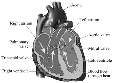
The atria and ventricles work in coordination. When the ventricles relax and fill with blood, the atria contract; and when the atria relax and fill, the ventricles contract and pump the blood forward — to the aorta on the left side and to the pulmonary arteries on the right side. The aorta is the largest artery in the body, originating at the exit of the heart, and from the aorta, all the other arteries branch off to provide blood to all the organs and tissues in the body. The pulmonary arteries carry the venous blood from the right side of the heart to the lungs to get oxygen and return this blood to the left side of the heart through the pulmonary veins.
The ventricles are the real powerhouses of the heart, particularly the left ventricle. If you look at the ventricles, you will see the thick muscle that makes up this chamber. In a normal heart, the left ventricle is more muscular than the right. Nonetheless, both ventricles handle the same volume of blood, so the right side of the heart is just as important as the left side.
The left and right sides of the heart are separate, meaning they do not exchange any blood directly across the atria or ventricles. If they do transfer blood between any of these chambers, there is likely a hole in the heart and this is abnormal. Normally, the heart keeps the venous blood (blood that has been depleted of its oxygen) from the right side of the heart separate from the oxygenated blood in the left side of the heart.
Did you know . . .
The heart beats approximately
♥ 70 beats a minute
♥ 10,000 beats a day
♥ 38 million beats a year
♥ 2.5 billion times over 70 years
The atria fill with blood from the venous systems that empty on both sides of the heart, but the right side of the heart gets the blood that has had most of its oxygen extracted and is returning from the body. The right side of the heart pumps the blood through the pulmonary artery to get oxygen from the lungs, and it returns the blood to the left side of the heart through the pulmonary veins, where the blood is now rich with oxygen. The left side of the heart pumps the blood through the aorta. This blood, now rich in oxygen, goes to all of the arteries in the body, including those that supply the heart muscle. You can understand how if the heart can’t work, meaning if it can’t pump blood to the body, the rest of the body can’t work. We need our hearts to function, otherwise all the other organs of the body will fail to get blood and, therefore, the oxygen they need to do their jobs. Ultimately this will cause all the organs to shut down and stop working. This is why the heart is the most important organ in the body. Without a working heart, we cannot live.
The Coronary Arteries
The coronary arteries provide the blood supply to the heart. These arteries get their blood supply every time the heart beats. Each time the left ventricle pumps blood forward through the aorta, the coronary arteries fill and provide the muscle of the heart with oxygen-rich blood so that the heart can work.
What Can Go Wrong with the Heart Arteries?
The heart muscle requires a constant supply of oxygen to function and this oxygen comes through the heart arteries. If the heart arteries are blocked, and thus the blood flow to the heart muscle is blocked or severely reduced, oxygen will not get to the heart muscle and the muscle can be damaged. This is what occurs when someone has a heart attack — also known as a myocardial infarction or, if you watch television medical dramas, an “MI.” (We discuss heart attacks in more detail in Chapter 6.) A myocardial infarction is the most common cause of heart disease in women, so we really want you to understand exactly how this occurs and how you can prevent it and treat it.
The coronary arteries can become blocked over time due to a gradual buildup of plaque, made up primarily of cholesterol. This process is known as atherosclerosis. If the cholesterol plaque ruptures or breaks in response to stress, a blood clot can form and block the artery completely, preventing blood from flowing forward to the area beyond the blockage. Since the heart muscle past the blockage is dependent on that artery for its oxygen supply, a blockage in blood flow will damage it. This condition of the heart muscle not receiving enough oxygen to function properly is known as ischemia. If the blood flow is not quickly restored, that affected area of the heart muscle may die (infarct).
Less common problems with the coronary arteries may be the result of birth defects in the heart artery anatomy, for example, congenital abnormalities in the positions of the coronary arteries. Such an abnormality may require surgical correction if the artery position results in compression of the artery, which would ultimately lead to ischemia, even when there is no plaque buildup. Vasospasm, where the heart artery contracts and reduces the blood flow to the heart muscle, is another problem.
The Heart Valves
The heart’s four valves keep the blood flowing in the appropriate direction. These valves are made up of leaflets (triangular-like flaps). The heart valves open and close within the heart cycle, allowing the blood to flow in one direction only: forward. When the valves are working normally they open and close fully and at appropriate times when the heart contracts. The heart valves between the atria and ventricles are known as the tricuspid valve and mitral valve, on the right and left side of the heart, respectively; the valve between the right ventricle and the pulmonary artery is the pulmonic valve; and the valve between the left ventricle and the aorta is called the aortic valve. Refer to the diagram of the heart on page 9 to see the position of the heart valves.
What Can Go Wrong with Heart Valves?
Two things can potentially go wrong with heart valves. First, they can leak and allow blood to flow backwards. This is known as regurgitation and can occur with any of the four valves. Second, the valves can become tight and narrow, making it difficult for blood to flow forward. This is known as stenosis, and it, too, can affect any of the four valves. In addition, more than one valve can be affected by the same disease process, and a person can have a leaky valve and tight valve at the same time.
Regurgitation and stenosis may be caused by congenital valve problems (that is, you are born with them), infections of the heart valves, rheumatic fever affecting the heart valves, damage to a heart valve or its structure after a heart attack or after developing heart failure, or valve disease from aging where there is calcification on the valves.
Symptoms are important signals, but changes to the heart can occur even without symptoms. If the valve is not repaired or replaced, more damage to the heart may occur, depending on how much the disease process affects the heart valves’ ability to open or close (or both) properly. Heart function can start declining as a result of the valve problem and, if left untreated, maybe irreversible.
The Heart Ventricles
The ventricles of the heart are in charge of pumping the nutrient-rich blood to the body and its organs, providing adequate nourishment and oxygen to all the organs so that they can function normally.
What Can Go Wrong with the Heart Ventricles?
When the ventricles of the heart are not working normally, the heart cannot pump blood as effectively as it should. When our heart cannot meet the body’s demands, we are said to have heart failure. Even though the term may imply that the heart can’t work at all, it can — just not as well as a healthy heart. There is a remarkable spectrum of severity of this disease. Some people with heart failure have no symptoms at all, while others are so limited by their heart failure that the simple act of moving about their home can be difficult. Heart failure can occur even when the heart function appears normal but the heart cannot relax normally; this is known as diastolic heart failure and is common in women.
Heart failure can result from several causes, including a heart attack or coronary artery disease (where one or more of the coronary arteries is narrowed), hypertension, valve disorders, chemotherapy drugs, alcohol, or infections or disease processes that affect the heart muscle.
Each year, 267,000 U.S. women die from heart attacks, which kill six times as many women as breast cancer. Another 31, 837 women die each year of congestive heart failure, representing 62.6% of all heart failure deaths.
The Electrical System of the Heart
What is really fascinating about the heart is that it has its own electrical system that responds to your body’s demands and requirements, telling the heart when to beat faster or slower, depending on how active you are. If you suddenly break into a run, your heart knows: your heart rate picks up. When you stop running, it knows it can safely and gradually slow down. When you sleep, the body’s demands for blood and oxygen lessen, so the heart slows down its rate even further from your resting heart rate of your waking hours.
It is the sinoatrial node (SA node or sinus node) located in the right atrium that usually controls the heart rate. When working properly, the SA node generates an electrical signal that then moves from the right atrium to the left atrium and on to the atrioventricular node (AV node), where through a specialized conduction system (His-Purkinje system) the electrical impulse is transferred to the ventricles. It is this electrical impulse that causes the heart to pump. If it is disrupted anywhere along this pathway, the heart rhythm may be affected, as well as the normal functioning of the heart.
If for some reason the sinoatrial node fails, many other places in the heart can take over. Basically, every cell that makes up the heart muscle is capable of sending a charge or signal to help keep the heart beating. Which part of the heart takes control determines the heart rate.
What Can Go Wrong with the Heart’s Electrical System?
Arrhythmias, irregular or abnormal heartbeats, are quite common and can range from inconsequential to life-threatening. Arrhythmias are more common as we age. We discuss in more detail the different rhythm disorders later, but briefly, your heart can beat too quickly (tachycardia), too slowly (bradycardia), or too early (either premature atrial contractions or PACs, or premature ventricular contractions or PVCs), which can feel like a skipped beat. Arrhythmias can be regular or irregular. Some arrhythmias may occur unnoticed — you only learn about them through a doctor’s exam or when an electrocardiogram is performed. Some arrhythmias require no treatment at all, some require medications, some require a pacemaker to regulate the heart rate, and some require immediate defibrillation to shock the heart to bring about a more stable rhythm. Some arrhythmias require an implanted defibrillator that will shock the heart when it detects that a dangerous arrhythmia is occurring.
Cardiac arrest, when the...
Table of contents
- Cover
- Title Page
- Table of Contents
- Dedication
- Acknowledgements
- Introduction
- The Heart Questions—Pompano Beach Heart Survey
- Chapter 1 - Affairs of a Woman’s Heart
- Chapter 2 - The Risks to a Woman’s Heart
- Chapter 3 - Determining Your Risk of Heart Disease
- Chapter 4 - Screening and Testing for Heart Disease: What Must Be Done, What Can Be Done, and What the Results Mean
- Chapter 5 - Heart Medications
- Chapter 6 - Therapeutic Options and Treatment Goals for Heart Disease and Specific Cardiac Risk Factors
- Chapter 7 - Nature’s Pharmacy
- Chapter 8 - Heart Bites
- Chapter 9 - Exercise and Your Heart
- Chapter 10 - The Impact of Stress
- Chapter 11 - Sleep and Heart Health
- Chapter 12 - Why Women Matter: Marching to a New Beat
- Resources
- Praise
- Endnotes
- Copyright
- About the Publisher
Frequently asked questions
Yes, you can cancel anytime from the Subscription tab in your account settings on the Perlego website. Your subscription will stay active until the end of your current billing period. Learn how to cancel your subscription
No, books cannot be downloaded as external files, such as PDFs, for use outside of Perlego. However, you can download books within the Perlego app for offline reading on mobile or tablet. Learn how to download books offline
Perlego offers two plans: Essential and Complete
- Essential is ideal for learners and professionals who enjoy exploring a wide range of subjects. Access the Essential Library with 800,000+ trusted titles and best-sellers across business, personal growth, and the humanities. Includes unlimited reading time and Standard Read Aloud voice.
- Complete: Perfect for advanced learners and researchers needing full, unrestricted access. Unlock 1.5M+ books across hundreds of subjects, including academic and specialized titles. The Complete Plan also includes advanced features like Premium Read Aloud and Research Assistant.
We are an online textbook subscription service, where you can get access to an entire online library for less than the price of a single book per month. With over 1.5 million books across 990+ topics, we’ve got you covered! Learn about our mission
Look out for the read-aloud symbol on your next book to see if you can listen to it. The read-aloud tool reads text aloud for you, highlighting the text as it is being read. You can pause it, speed it up and slow it down. Learn more about Read Aloud
Yes! You can use the Perlego app on both iOS and Android devices to read anytime, anywhere — even offline. Perfect for commutes or when you’re on the go.
Please note we cannot support devices running on iOS 13 and Android 7 or earlier. Learn more about using the app
Please note we cannot support devices running on iOS 13 and Android 7 or earlier. Learn more about using the app
Yes, you can access Saving Women's Hearts by Martha Gulati,Sherry Torkos in PDF and/or ePUB format, as well as other popular books in Medicine & General Health. We have over 1.5 million books available in our catalogue for you to explore.