The human body includes very effective and efficient technology, such as light receptors (eyes), chemical receptors (tongue and nose), and movement (muscles). This book explains how these functions work on the molecular level and then discusses nanotechnology that uses the same structure-function relationships.
- 114 pages
- English
- ePUB (mobile friendly)
- Available on iOS & Android
eBook - ePub
About this book
Trusted by 375,005 students
Access to over 1.5 million titles for a fair monthly price.
Study more efficiently using our study tools.
Information
1Introduction
This book is about human senses and movement, how they function on the nanoscale, and how they can be mimicked on the nanoscale by technology. To understand this subject, it is necessary to first know a little about what happens in the human body and what takes place at the nanoscale more generally. This introduction will help you by providing an overview on several topics. It will also include common measurement techniques and an introduction to sensors. Feel free to only look at the sections you need help with; each section should be understandable by itself. References will be provided if you want to look into a topic in more detail.
1.1Introduction to Cell Biology and Cell Communication
Let’s start with some basic physiology [1]. Skin, the biggest organ of the body, surrounds and protects the other organs, each with their separate function. The skeleton holds up all of the organs and cells, the brain coordinates reactions to our environment, plans and regulates movement, the lungs provide oxygen so that fuel can be burnt and energy for the body created, this fuel is collected and broken down in the stomach, but toxic compounds and waste are secreted via the kidneys. The liver produces whatever needs to be produced to make it all work, and the heart is responsible for the transport of nutrients throughout the body. Why does each function have its own organ?
The short answer is that for each organ to be able to function correctly it needs its own environment [1]. Each organ has different genes that are active and works with different chemical compounds in its cells, using a variety of reactions to reach its goals. And each organ, and the body itself, can only function in very controlled conditions within a narrow margin. These narrow margins are called homeostasis. The body has an overall homeostasis or set of requirements and conditions, but so does each organ and each individual cell.
To maintain those conditions, membranes envelop the body, each organ, and each cell. These border-enclosed spaces can then be controlled separately. But here is the problem with this approach: each of those separated spaces, including each cell, must still work together to create a functioning body. Humans have trillions and trillions of cells, and yet humans are not just a random heap of cells. The cells organize themselves into tissues, each tissue is ordered into organs, and each organ is combined to form a human. Humans have a hard time organizing several people; how can the body organize trillions of cells? And not only organize the cells into a static structure, but create an active, environment-responsive, moving and thinking human? The answer is: communication, communication, communication.
Before we get to the communication part, let us look at the structure of a cell and how it functions (Figure 1.1). The nucleus contains all information for the cell coded in the double-stranded deoxyribonucleic acid (DNA). The sequence of four different bases in DNA gets translated into ribonucleic acid (RNA) and then proteins by ribosomes, and the proteins are distributed to their specific locations in the cell via the endoplasmic reticulum (ER) and the Golgi complex. Some proteins are structural, creating the different structures in the cell and human body. Most of the proteins are enzymes, i.e. they catalyze reactions and thus either construct needed compounds in the cell or break them down to generate energy (anabolism or catabolism, respectively). Some catalysts are also part of signal transduction pathways and thus part of cell communication.

The place where energy is generated is in the mitochondria. Lysosomes, vacuoles, and peroxisomes either ingest fuel or break down toxic compounds. The cytoskeleton is made from structural protein fibers and holds the shape of the cell and act as “roads” for organelle and other transport throughout the cell. The cell membrane is the barrier that allows for homeostasis. It contains a lot of proteins which are mostly channels that allow for very specific and regulated transport in and out of the cell. Other transport across the membranes operates via vesicles that are endocytosed or exocytosed. Other transmembrane proteins are designed to transfer signals. Some proteins on the outside of the cell membrane act as a “marker”, or recognition element, for the cell, and some cause the cell to adhere to other cells or the extracellular matrix (ECM). ECM consists of fibers (collagen, elastin) and an amorphous matrix (proteoglycans, cell-binding adhesive glycoproteins, solutes, and water) which together form a cell-support. The mechanical strength of the tissue depends on both the strength of the cytoskeleton for each cell and the strength of the ECM. The ordering of cells into tissues is also regulated by the ECM.
Cells are not static structures but grow, develop and mature, and multiply. They also react to stimuli, outside forces and conditions, and might even commit suicide (apoptosis) if they are irreversibly damaged or are infected with a dangerous virus or bacterium. The centriole starts the process of cell division (mitosis) by making the spindle fibers that draw the two strands of the chromosomes (DNA pieces) apart. The cells can then form two nuclei, one for each cell, by copying the one strand, reforming the complete, double-stranded DNA chromosome and then forming a nuclear membrane around it; after that, the complete cell divides by forming a membrane between those nuclei and separating the identical cells from each other.
To modify a cell, specifically to modify or remove a specific protein in a cell, the method of genetic engineering was developed. It has been most widely used in bacteria and plants. The genetic information for a specific protein, its gene or piece of DNA sequence, is excised and modified. To be active in the cell, that piece of DNA needs to be attached to a promoter sequence that regulates when and how that information is used, then the DNA is reinserted into the cell. This method works most effectively if one function is based on only one gene, which is common in simple organisms such as bacteria but rare for higher organisms.
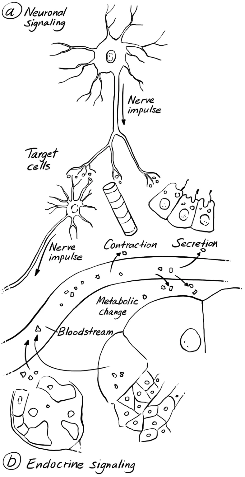
With this information on cell structure in mind, let us return to cell communication. The body has two major communication systems: the endocrine system that uses hormones as its signal and the neuronal system that uses neurotransmitters instead (Figure 1.2). Hormones are secreted into the blood stream and are widely distributed. They act on any tissue that happens to have receptors for that specific hormone. Hormones are generally signals for development, i.e., slower, more long-term processes such as the signal for a stem cell to develop into a new neuron in the brain, or signals that activate several organs, i.e. insulin, which activates several organs so that food can be broken down and converted into energy.
Neurotransmitters will be released into a very small space between two nerve cells and thus will act only on that subsequent neuron. Release and uptake are fast, and the neurotransmitter will be destroyed immediately. So these signals are used to react quickly to the environment or analyze and transmit information in the brain stemming from the continuous input of our senses.
In either communication system, though, the signal arrives outside of the cell that is supposed to use that information. So how does the signal on the outside of the cell effect a change inside of that cell, given that the cell membrane poses such a formidable barrier? Here, the specific channels or other transmembrane proteins come into play. The structures and mechanisms of these channels will be discussed in more detail in the next chapter (see Section 1.2). The signal binds to a channel or a transmembrane protein on the outside of the cell. That binding event changes the three-dimensional structure of that protein so that an enzyme at the inside of the cells gets activated to catalyze a reaction. The product of that reaction then binds to another enzyme, which activates it to perform another reaction, whose product binds and activates another enzyme, and so on. This process is called a signal cascade.
Why does a signal cascade take place, instead of just one single reaction? Doesn’t such a complex process create a greater risk of something going wrong? In fact, the opposite is the case: Each of the steps in the signal cascade can be regulated, thus allowing the signal effect to be tightly controlled. Also, a signal cascade creates many signals for a lot of proteins in a short time, and thus creates signal amplification, which allows for fast, coordinated action. For example, within a muscle, a large number of heads of myosin have to be activated at the same time, otherwise only a few muscle fibers would contract instead of the complete muscle.
We will look at two common signal transduction pathways as examples. The first is the adenylate cyclase pathway (Figure 1.3). The transmembrane receptor is associated with a G-protein (a protein that hydrolyses guanine triphosphate (GTP) to guanosine diphosphate (GDP)). These complexes are called G-protein-coupled receptors, or GPCR. The G-protein is phosphorylated and in turn phosphorylates the enzyme adenylate cyclase. The thus activated adenylate cyclase takes adenylate monophosphate (AMP) and cyclizes it to cAMP; cAMP is an important signal in the cell that can bind and activate many kinases. Kinases are enzymes that can phosphorylate other enzymes and thus activate or inactivate enzymes, thereby allowing or barring specific reactions. Each of the enzymes in the pathway can be regulated either by binding a different compound into the active site of the enzy...
Table of contents
- Cover
- Title Page
- Copyright
- Preface
- Contents
- List of abbreviations
- 1 Introduction
- 2 Movement
- 3 Vision
- 4 Smell and Taste
- 5 Hearing
- 6 Skin, The Body’s Largest Organ
- 7 Future Developments
- Index
Frequently asked questions
Yes, you can cancel anytime from the Subscription tab in your account settings on the Perlego website. Your subscription will stay active until the end of your current billing period. Learn how to cancel your subscription
No, books cannot be downloaded as external files, such as PDFs, for use outside of Perlego. However, you can download books within the Perlego app for offline reading on mobile or tablet. Learn how to download books offline
Perlego offers two plans: Essential and Complete
- Essential is ideal for learners and professionals who enjoy exploring a wide range of subjects. Access the Essential Library with 800,000+ trusted titles and best-sellers across business, personal growth, and the humanities. Includes unlimited reading time and Standard Read Aloud voice.
- Complete: Perfect for advanced learners and researchers needing full, unrestricted access. Unlock 1.5M+ books across hundreds of subjects, including academic and specialized titles. The Complete Plan also includes advanced features like Premium Read Aloud and Research Assistant.
We are an online textbook subscription service, where you can get access to an entire online library for less than the price of a single book per month. With over 1.5 million books across 990+ topics, we’ve got you covered! Learn about our mission
Look out for the read-aloud symbol on your next book to see if you can listen to it. The read-aloud tool reads text aloud for you, highlighting the text as it is being read. You can pause it, speed it up and slow it down. Learn more about Read Aloud
Yes! You can use the Perlego app on both iOS and Android devices to read anytime, anywhere — even offline. Perfect for commutes or when you’re on the go.
Please note we cannot support devices running on iOS 13 and Android 7 or earlier. Learn more about using the app
Please note we cannot support devices running on iOS 13 and Android 7 or earlier. Learn more about using the app
Yes, you can access Biomimetic Nanotechnology by Anja Mueller in PDF and/or ePUB format, as well as other popular books in Technology & Engineering & Biochemistry. We have over 1.5 million books available in our catalogue for you to explore.