Anaphylaxis is the most dramatic and potentially life-threatening manifestation of an immediate-type hypersensitivity reaction. Although known for over 100 years, it still poses many unresolved questions, and its practical management and acute treatment are often more empiric in nature than evidence-based.In this book a multidisciplinary group of experts review the state of the art in the pathophysiology, epidemiology, diagnosis and clinical symptomatology of anaphylaxis. Its etiology with regard to different elicitors such as insect venoms, radiocontrast media, analgesics, general and local anesthetics is examined in detail. Finally, treatment modalities for anaphylaxis are discussed both for acute reactions and as general management recommendations for patients at risk. Providing thorough and up-to-date coverage of this frequently underestimated problem, this book is of interest not only to allergologists and immunologists, but also to all physicians and affected patients.
- 254 pages
- English
- ePUB (mobile friendly)
- Available on iOS & Android
eBook - ePub
Anaphylaxis
About this book
Trusted by 375,005 students
Access to over 1.5 million titles for a fair monthly price.
Study more efficiently using our study tools.
Information
Topic
MedicineSubtopic
Anesthesiology & Pain ManagementMechanisms
Ring J (ed): Anaphylaxis. Chem Immunol Allergy. Basel, Karger, 2010, vol 95, pp 22–44
______________________
T-Cell Response to Allergens
Cevdet Ozdemira · Mübeccel Akdisb · Cezmi A. Akdisb
aMarmara University, Division of Pediatric Allergy and Immunology, Istanbul, Turkey and bSwiss Institute of Allergy and Asthma Research (SIAF), Davos, Switzerland
______________________
Abstract
Anaphylaxis is a life-threatening IgE-dependent type 1 hypersensitivity reaction in which multiple organ systems are involved. The existence of allergen exposure and specific IgE are the major contributors to this systemic reaction. The decision of the immune system to respond to allergens is highly dependent on factors including the type and load of allergen, behavior and type of antigenpresenting cells, innate immune response stimulating substances in the same micromilieu, the tissue of exposure, interactions between T and B lymphocytes, costimulators, and genetic propensity known as atopy. Antigen-presenting cells introduce processed allergens to T-helper lymphocytes, where a decision of developing different types of T-cell immunity is given under the influence of several cytokines, chemokines, costimulatory signals and regulatory T cells. Among Th2-type cytokines, interleukin (IL)-4 and IL-13 are responsible for class switching in B cells, which results in production of allergen-specific IgE antibodies that bind to specific receptors on mast cells and basophils. After re-exposure to the sensitized allergen, this phase is followed by activation of IgE Fc receptors on mast cells and basophils resulting in biogenic mediator releases responsible for the symptoms and signs of anaphylaxis. Since the discovery of regulatory T cells, the concepts of immune regulation have substantially changed during the last decade. Peripheral T-cell tolerance is a key immunologic mechanism in healthy immune response to self antigens and non-infectious non-self antigens. Both naturally occurring CD4+CD25+ regulatory T (Treg) cells and inducible populations of allergenspecific, IL-10-secreting Treg type 1 cells inhibit allergen-specific effector cells and have been shown to play a central role in the maintenance of peripheral homeostasis and the establishment of controlled immune responses. On the other hand, Th17 cells are characterized by their IL-17 (or IL-17A), IL-17F, IL-6, tumor necrosis factor-α, and IL-22 expressions, which coordinate local tissue inflammation through upregulation of proinflammatory cytokines and chemokines. This chapter is mainly focused on antigen presentation pathways and allergen-specific T-cell responses.
Copyright © 2010 S. Karger AG, Basel
The terms ‘allergy’ and ‘atopy’are in close proximity of our lives in the new millennium since our lifestyles have enormously changed. Encounters with various new molecules in air, water and diet, living in a more polluted world with less exposure to infections, and infectious agents are supposed to be the major causative factors added to the genetic propensity of developing IgE antibodies responsible for symptoms and signs of allergic disorders [1]. Clinical manifestations are allergic rhinitis, allergic asthma, food allergy, allergic skin inflammation, ocular allergy as a single or combined disease and anaphylaxis [2].
Allergens are almost always proteins, but not all proteins are allergens. Understanding what makes a protein an allergen is essential to develop strategies for immune intervention [2]. For a protein antigen to display allergenic activity, it must induce IgE production, which must lead to a type 1 hypersensitivity response upon subsequent exposure to the same protein [3]. Biochemical properties of the allergen, stimulating factors of the innate immune response around the allergen substances at the time of exposure, stability of the allergen in the tissues, digestive system, skin or mucosa, and the dose and time of stay in lymphatic organs during the interaction with the immune system are all possible confounding factors causing an antigen to become an allergen [2]. Foods (especially peanuts and tree nuts), medications (also allergen immunotherapy injections), insect venoms and latex constitute the major allergens causing anaphylaxis [4, 5]. Early detection of the responsible allergen is a requisite and an important prognostic factor in controlling allergic diseases and anaphylaxis [6].
Dendritic cells (DCs) are complex cell populations that differ in their anatomic location, antigen recognition, processing machinery, and migratory capacity. DCs stay as sentinels that take up exogenous antigens and transmit the information into immune system by migrating to draining lymph nodes, and presenting the processed antigens to T cells resulting in T-cell differentiation and activation [7, 8]. Content of micromilieu and several cytokines and other cofactors released from DCs are essential for the differentiation of naive T cells into T-helper (Th)1, Th2, Th9, Th17 effector T-cell subsets [9]. Expansion of allergen-specific Th2 cells results in production of interleukin (IL)-4 and IL-13, which induce immunoglobulin class switching to IgE and clonal expansion of naive and IgE+ memory B-cell populations. In the presence of IL-4 also differentiation of naive T cells into Th2 takes place (fig. 1). When IgE bound to FcεRI (high-affinity receptor for IgE) on mast cells and basophils crosslinks with the specific allergen, release of vasoactive amines (such as histamine), lipid mediators (such as prostaglandin D, platelet-activating factor, leukotriene C4 (LTC4), LTD4 and LTE4), chemokines (CXC-chemokine ligand 8 (CXCL8), CXCL10, CC-chemokine ligand 2 (CCL2), CCL4 and CCL5) and other cytokines (such as IL-4, IL-5 and IL-13) occur, which are responsible for the signs and symptoms of immediate phase of the allergic reactions [3].
Allergen Recognition by the Immune System
Pathogen-Associated Molecular Patterns and Pattern Recognition Receptors
Recent investigations have greatly increased our understanding of immunological mechanisms involved in the pathogenesis of allergic disease [10-12]. Presentation of allergens by antigen-presenting cells (APCs) or by other means and initiation of allergen-specific immune response represents the first step in sensitization to allergens. New allergens and their cross-reactivities are continuously being identified and types of immune response to them are demonstrated [13, 14]. Allergic inflammation results from the activation of tissue migrating hematopoietic and resident non-hematopoietic cells. This coordinated activation leads to increased production of a variety of soluble factors including chemokines and cytokines. Direct or indirect effects of the innate immune response are decisive in the development of adoptive immunity to allergens [15]. In principle, it is not only the protein allergen, but the adjuvants in the surrounding of the allergens are decisive for the type of the immune response [16, 17].
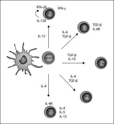
Fig.1. Differentiation of T cells: content of micromilieu and several cytokines and other cofactors released from DCs are essential for the differentiation of naive T cells into T-helper(Th)1, Th2, Th9, Th 17 effectorT-cell subsets.
Mammals execute host defense against pathogens through two different types of immunity: innate and adaptive immune responses [18]. APCs and lymphocytes act as important contributors in the imminent relationship between these two systems. Innate immunity is designed to recognize small molecular motifs that are unique and essential for the survival of pathogens, which do not exist in mammalians termed as pathogen-associated molecular patterns (PAMPs) [19-21]. PAMPs are recognized by pattern recognition receptors (PRRs) that are expressed by DCs. Among the well-known PRRs, TLRs are the best-characterized group that recognize bacteria or viruses [22, 23]. TLR3, TLR7, TLR8, and TLR9 recognize viral RNAs and bacterial DNA [24]. TLR3 is expressed on the surface of airway epithelial cells [25]. Moreover, TLR engagement on DCs polarizes T-cell response and while TLR2 and TLR4 may favor both Th1 and Th2 responses, and TLR9 induces the development of regulatory T cells [26]. Toll is a type I transmembrane receptor containing extracellular leucinerich repeat (LRR) motifs and cytoplasmic Toll/IL-1 receptor homology domain (TIR) [27]. TLRs activate nuclear factor (NF)-KB and other signaling pathways such as mitogen-associated protein (MAP) kinases, signal transducer and activator of transcription (STAT)-1 through the adapter protein MyD88, TIR containing adaptor protein (TIRAP), TIR containing adaptor inducing interferon (IFN)-β (TRIF) and TRIF-related adaptor molecule (TRAM) [26, 28-30].
PAMPs and various tissue factors can prime DCs to produce T-cell-polarizing factors [21]. IL-12 is a pro-inflammatory cytokine that induces IFN-γ and promotes the development of Th1-cell differentiation [31]. Other Th1-polarizing factors are IFN-α and IFN-β [32] and cell-surface expressed intracellular adhesion molecule (ICAM)-1 [33]. On the other hand, it has been shown that NF-KB inducing kinase (NIK), which is known to regulate B-cell maturation and lymphoid organogenesis, is important for the induction of Th17 cells [34].
TLR signals in DCs increase expression of major histocompatibility complex (MHC) proteins and T-cell coreceptors, resulting i...
Table of contents
- Cover Page
- Front Matter
- History and Classification of Anaphylaxis
- Epidemiology of Anaphylaxis
- T-Cell Response to Allergens
- Anaphylaxis: Mechanisms of Mast Cell Activation
- Kinins, Airway Obstruction, and Anaphylaxis
- Role for Basophils in Systemic Anaphylaxis
- Human Cardiac Mast Cells in Anaphylaxis
- Mastocytosis
- In vitro Diagnosis of Anaphylaxis
- Insect Venoms
- Classification and Pathophysiology of Radiocontrast Media Hypersensitivity
- Analgesics
- Anaphylaxis to General Anesthetics
- Anaphylactic Reactions to Local Anesthetics
- Anaphylaxis: Acute Treatment and Management
- Epinephrine (Adrenaline) in Anaphylaxis
- Author Index
- Subject Index
Frequently asked questions
Yes, you can cancel anytime from the Subscription tab in your account settings on the Perlego website. Your subscription will stay active until the end of your current billing period. Learn how to cancel your subscription
No, books cannot be downloaded as external files, such as PDFs, for use outside of Perlego. However, you can download books within the Perlego app for offline reading on mobile or tablet. Learn how to download books offline
We are an online textbook subscription service, where you can get access to an entire online library for less than the price of a single book per month. With over 1.5 million books across 990+ topics, we’ve got you covered! Learn about our mission
Look out for the read-aloud symbol on your next book to see if you can listen to it. The read-aloud tool reads text aloud for you, highlighting the text as it is being read. You can pause it, speed it up and slow it down. Learn more about Read Aloud
Yes! You can use the Perlego app on both iOS and Android devices to read anytime, anywhere — even offline. Perfect for commutes or when you’re on the go.
Please note we cannot support devices running on iOS 13 and Android 7 or earlier. Learn more about using the app
Please note we cannot support devices running on iOS 13 and Android 7 or earlier. Learn more about using the app
Yes, you can access Anaphylaxis by J. Ring, T. A. E. Platts-Mills in PDF and/or ePUB format, as well as other popular books in Medicine & Anesthesiology & Pain Management. We have over 1.5 million books available in our catalogue for you to explore.