In systemic childhood diseases, including cancer, gastrointestinal, pulmonary and cardiac disorders, childhood growth is severely impaired. In addition, almost 400 known genetic diseases inhibit the ability of the growth plate to form new bone, leaving affected children with growth failure and bony deformities which can severely impact their quality of life and may lead to morbidity and early mortality. This book provides a comprehensive review of bone and cartilage development, growth and disease. Focusing on novel treatment strategies, regulatory signals and molecular mechanisms are discussed in relation to the diseases affecting them. Furthermore, novel methodologies in bone and cartilage research based on recent advances in skeletal stem cell biology, cartilage tissue engineering and allele-specific gene silencing is covered. Providing insight into the basic mechanisms of bone growth, structure and metabolism, research methodology, as well as discussing the clinical management of related diseases, this book is of particular value to physicians with a special interest in bone and cartilage biology; in particular endocrinologists and pediatric endocrinologists that see patients with growth disorders, osteoporosis, osteogenesis imperfecta, and skeletal dysplasias.
eBook - ePub
Cartilage and Bone Development and Its Disorders
4th ESPE Advanced Seminar in Developmental Endocrinology, Stockholm, June-July 2010
- 132 pages
- English
- ePUB (mobile friendly)
- Available on iOS & Android
eBook - ePub
Cartilage and Bone Development and Its Disorders
4th ESPE Advanced Seminar in Developmental Endocrinology, Stockholm, June-July 2010
About this book
Information
Topic
MedizinCamacho-Hübner C, Nilsson O, Sävendahl L (eds): Cartilage and Bone Development and Its Disorders.
Endocr Dev. Basel, Karger, 2011, vol 21, pp 1-11
Endocr Dev. Basel, Karger, 2011, vol 21, pp 1-11
______________________
Chondrocyte Proliferation and Differentiation
Manuela Wuelling · Andrea Vortkamp
Center for Medical Biotechnology, Department of Developmental Biology, University Duisburg- Essen, Essen, Germany
______________________
Abstract
The skeletal elements of the axial and appendicular skeleton are preformed as cartilage templates by a mechanism called endochondral ossification. During this process, a cartilage template is formed in which chondrocytes proliferate and differentiate into hypertrophic chondrocytes and are gradually replaced by bone. Postnatally, remnants of embryonic chondrocytes remain in a restricted domain between the ossified regions of the bones forming the growth plate. The coordinated proliferation and differentiation of chondrocytes ensures the continuous elongation of the epiphyseal growth plates. The sequential changes between proliferation and differentiation are tightly regulated by secreted growth factors, which activate chondrocyte-specific transcription factors. Transcription factors that play critical roles in regulating cell type-specific gene expression include SOX9, GLI2/3 and RUNX2. This review will outline recent advances in the analysis of the complex transcriptional network that regulates distinct steps of chondrocyte differentiation.
Copyright © 2011 S. Karger AG, Basel
Endochondral Ossification
During endochondral ossification, bones are formed by two processes. During intramembranous ossification, by which the bones of the skull and part of the facial bones and clavicle are formed, mesenchymal cells directly differentiate into osteoblasts. In contrast, the axial and appendicular skeleton of vertebrates develops by endochondral ossification. During this process, the initial anlagen are formed as cartilage templates, which model the skeletal elements. Gradually, the cartilage templates are replaced by bone. The process of endochondral ossification starts with the condensation of mesenchymal cells, which differentiate into chondrocytes (fig. 1a, b). These cells initially proliferate and differentiate into two subpopulations of proliferating chondrocytes: round, low proliferating chondrocytes at the distal ends of the condensation (RP chondrocytes) and high proliferating chondrocytes that are organized in columns (CP chondrocytes) towards the center [1–3]. In the center of the growing cartilage anlagen, chondrocytes exit the cell cycle and differentiate into prehypertrophic and hypertrophic chondrocytes (fig. 1c, 2a). These produce a mineralized extracellular cartilage matrix that serves as a template for the subsequent deposition of bone matrix. The increasing volume of the hypertrophic cells contributes significantly to the elongation of the skeletal elements.
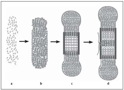
Fig. 1. Endochondral ossification requires sequential differentiation and degradation of chondrocytes. Mesenchymal cells condense to form the cartilage anlagen (a) and differentiate into proliferating chondrocytes and fibroblast-like cells in the perichondrium (b). c Chondrocytes form distinct subpopulations, which can be morphologically distinguished from distal to central into RP and CP chondrocytes. After cell cycle exit, chondrocytes differentiate into prehypertrophic, hypertrophic and terminal hypertrophic cells. Adjacent to the hypertrophic zone, cells in the perichondrium mineralize and form the periosteum. d Blood vessels invade the hypertrophic region and initiate bone formation in the center of the skeletal elements.
The cartilage elements are surrounded by a layer of fibroblast-like cells, the perichondrium. At stages when chondrocytes become hypertrophic, cells in the perichondrium differentiate into osteoblasts forming the periost, which is highly vascularized. Blood vessels from the periost invade the calcified matrix of terminal hypertrophic chondrocytes, which undergo apoptosis. Together with the blood vessels osteoblasts and osteoclasts invade the skeletal elements to replace the mineralized cartilage by bone generating the primary spongiosa (fig. 1c, d) [4, 5]. In parallel, osteoblasts in the periost produce a calcified matrix forming the bone collar. The early bone collar and the primary trabecles are later remodeled to form the cortical bone and the bone marrow cavity, which provides a local environment for hematopoiesis.
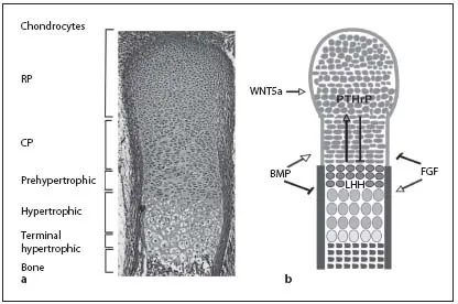
Fig. 2. In the embryonic growth plates, distinct chondrocyte populations can be morphologically distinguished. a Safranin-Weigert staining of a longitudinal section through an E16.5 mouse forelimb showing the characteristic morphology of the cartilage. b Schematic model of a longitudinal section through an embryonic growth plate showing the different chondrocyte subpopulations. Growth factors (BMP, FGF, IHH, PTHrP and WNT5a) can activate (arrows) or inhibit (bars) the formation of distinct chondrocyte populations.
During postnatal development, secondary ossification centers are formed within the zone of the RP chondrocytes (fig. 1d). Between the two ossification centers chondrocytes remain forming the postnatal growth plate. Many studies investigating postnatal growth plate differentiation strongly indicate that the mechanisms controlling the embryonic processes are maintained until adulthood [6].
Longitudinal growth of bones is dependent on the rate of chondrocyte proliferation, the size of the proliferating region and the expansion of the hypertrophic region. During embryonic and postnatal development, the balance between chondrocyte proliferation and differentiation has to be tightly controlled to ensure normal longitudinal bone growth and endochondral bone formation. Both, disturbed chondrocyte proliferation and hypertrophic differentiation, will lead to alterations in bone length and stability.
The sensitivity of the system is reflected by the large number of human chondrodysplasias and the multitude of transgenic mice that show skeletal defects [7]. During the last years, considerable progress has been made in analyzing the signals regulating endochondral ossification. However, the mechanisms by which these signals are translated into gene expression are less well understood. Here, we will summarize some of the relevant signaling pathways and concentrate on recent advances in deciphering the transcriptional network acting downstream of these pathways.
Regulation by Growth Factors
Indian Hedgehog Signaling
One of the key regulators of endochondral bone formation is the secreted growth factor IHH which is expressed in prehypertophic chondrocytes. IHH signals to the proliferating chondrocytes and to the periosteum. In proliferating chondrocytes, IHH signaling directly activates proliferation, especially in CP chondrocytes. In parallel, high IHH signals repress the expansion of the pool of RP chondrocytes, whereas low IHH levels favor its expansion. Furthermore, IHH induces the expression of PTHrP in RP chondrocytes. Activation of the PPR1, expressed in proliferating and prehypertrophic chondrocytes, by PTHrP prevents the onset of hypertrophic differentiation and, thus, the differentiation of IHH-expressing cells (fig. 2b) [8, 9]. By initiating this negative feedback system, IHH regulates the pool of proliferating cells in parallel to the proliferation rate.
Signaling of IHH is mediated by transcription factors of the GLI family, which consist of GLI1, GLI2 and GLI3. GLI1 activates IHH target genes, while GLI2 and GLI3 can function as activators or can be processed into a repressor form [10, 11]. This proteolytic processing is negatively regulated by IHH signaling leading to distinct levels of GLI activator and GLI repressor forms in different chondrocyte subpopulations.
Bone Morphogenetic Protein Signaling
BMPs were identified by their ability to induce ectopic endochondral ossification. Several members of the BMP family, which represents a subgroup of the transforming growth factor family of secreted proteins, and their receptors (BMPRs) are expressed in the cartilage anlagen [12]. BMP signaling is mediated by heteromeric complexes of type I (BM PR1a and BMPR1b) and type II (BMPR2) receptors. Activation of these receptor complexes leads to the phosphorylation of SMAD transcription factors SMAD1, SMAD5 and SMAD8. Phosphorylated SMAD proteins interact with the common mediator SMAD4 to activate transcription of target genes [13]. BMP signaling induces chondrocyte proliferation, whereas loss of both BMPR1a and 1b leads to hypoplastic cartilage anlagen, identifying BMPs as positive regulators of cell cycle progression [4, 14, 15].
Fibroblast Growth Factor Signaling
Like BMPs, FGFs are members of a large family of signaling proteins, which act at multiple steps of the differentiation process. FGF signaling leads to activation of the MAP pathway and phosphorylatio...
Table of contents
- Cover Page
- Front Matter
- Chondrocyte Proliferation and Differentiation
- Local Regulation of Growth Plate Cartilage
- Growth Plate Senescence and Catch-Up Growth
- Endocrine Regulation of Longitudinal Bone Growth
- Estrogen Signaling in Growth Plate Cartilage
- Methods to Study Cartilage and Bone Development
- Fibroblast Growth Factor-23 and Phosphorus Metabolism
- Molecular Defects Causing Skeletal Dysplasias
- Allele-Specific Gene Silencing in Osteogenesis Imperfecta
- Stem Cells in Skeletal Physiology and Endocrine Diseases of Bone
- Cartilage Tissue Engineering
- Author Index
- Subject Index
Trusted by 375,005 students
Access to over 1.5 million titles for a fair monthly price.
Study more efficiently using our study tools.
Frequently asked questions
Yes, you can cancel anytime from the Subscription tab in your account settings on the Perlego website. Your subscription will stay active until the end of your current billing period. Learn how to cancel your subscription
No, books cannot be downloaded as external files, such as PDFs, for use outside of Perlego. However, you can download books within the Perlego app for offline reading on mobile or tablet. Learn how to download books offline
Perlego offers two plans: Essential and Complete
- Essential is ideal for learners and professionals who enjoy exploring a wide range of subjects. Access the Essential Library with 800,000+ trusted titles and best-sellers across business, personal growth, and the humanities. Includes unlimited reading time and Standard Read Aloud voice.
- Complete: Perfect for advanced learners and researchers needing full, unrestricted access. Unlock 1.5M+ books across hundreds of subjects, including academic and specialized titles. The Complete Plan also includes advanced features like Premium Read Aloud and Research Assistant.
We are an online textbook subscription service, where you can get access to an entire online library for less than the price of a single book per month. With over 1.5 million books across 990+ topics, we’ve got you covered! Learn about our mission
Look out for the read-aloud symbol on your next book to see if you can listen to it. The read-aloud tool reads text aloud for you, highlighting the text as it is being read. You can pause it, speed it up and slow it down. Learn more about Read Aloud
Yes! You can use the Perlego app on both iOS and Android devices to read anytime, anywhere — even offline. Perfect for commutes or when you’re on the go.
Please note we cannot support devices running on iOS 13 and Android 7 or earlier. Learn more about using the app
Please note we cannot support devices running on iOS 13 and Android 7 or earlier. Learn more about using the app
Yes, you can access Cartilage and Bone Development and Its Disorders by C. Camacho-Hübner,O. Nilsson,L. Sävendahl, C. Camacho-Hübner, O. Nilsson, L. Sävendahl in PDF and/or ePUB format, as well as other popular books in Medizin & Alternativ- & Komplementärmedizin. We have over 1.5 million books available in our catalogue for you to explore.