eBook - ePub
Histopathology of Chronic Constipation
- 64 pages
- English
- ePUB (mobile friendly)
- Available on iOS & Android
eBook - ePub
Histopathology of Chronic Constipation
About this book
The symptom of chronic constipation is often caused by a series of intestinal diseases, which can be reliably diagnosed histopathologically by histochemical techniques and consequently treated by surgical intervention. The following publication is the second and completely revised edition of 'Pathology of Chronic Constipation in Pediatric and Adult Coloproctology' published in 2005, and introduces several new diseases and figures. It includes characteristics of classical and ultrashort Hirschsprung's disease as well as total intestinal aganglionosis and hypoganglionosis. New diseases such as intestinal neuronal dysplasia, desmosis coli, leiomyopathy, architectural malformation, and stretching lesions of muscularis propria are critically discussed. Atrophic desmosis is also covered. This new and frequently observed degeneration of muscularis propria in Crohn's disease, sigmoid diverticulitis, and other inflammatory intestinal diseases causes focal aperistalsis, frequently interpreted as scar stenosis. 'Histopathology of Chronic Constipation' provides a comprehensive overview of intestinal alterations which cause chronic constipation. It is therefore of special interest to diagnostic pathologists, clinicians, pediatric and abdominal surgeons, coloproctologists, and gastroenterologists.
Trusted by 375,005 students
Access to over 1.5 million titles for a fair monthly price.
Study more efficiently using our study tools.
Information
Topic
MedicineSubtopic
Gastroenterology & Hepatology
Different Colon Diseases with Chronic Constipation
B.1
Pathogenesis of Hirschsprung’s Disease
Neuroblasts of the neck vagus migrate during embryonic weeks 6-12 craniocaudally along circular muscles to form a myenteric plexus. With a delay of several days, nerve cells of the myenteric plexus migrate into the submucosa to generate a submucous plexus.
During embryonic weeks 5-12, nerve fibers invade into circular muscles and mucosa of the distal colon from the sacral roots S2-S5. This happens 4 to 5 weeks before neuroblasts arrive in the descending colon. These nerves from the sacrum release acetylcholine in a synchronous manner. This causes a spastic contraction in the rectosigmoid up to the synaptic linking of these nerves with the invading neurocrest cells. This is a long-lasting process from embryonic weeks 10-12. It is a very sensitive period during which neuroblasts connect synaptic with parasympathetic nerves of sacral roots S2-S5. If neuroblasts do not arrive in the distal colon, HD or an aganglionosis develops. Therefore, HD is limited to the rectosigmoid or rectum in about 75% of babies.
In cases of rectum aganglionosis, nerve cells which modulate the permanent firing nerves from the sacral roots are missing, causing a spastic contraction of circular muscles and thus gut obstruction. Because acetylcholine and AChE have the same level, AChE can be used to
evaluate the cholinergic level of a particular tissue. This fact is used in enzyme histochemistry to recognize an aganglionosis in the rectal mucosa. The increase of AChE activity in parasympathetic nerve fibers of the rectum mucosa can be used as a reliable indicator of an aganglionosis. AChE activity increases dependently with age (fig. 2-6). LDH and SDH or nitroxide synthase (NOS), which stain nerve cells electively, can be used as a second indicator of an aganglionosis (fig. 7).
Indicators of Hirschsprung’s Disease
1 Increased acetylcholine release and AChE activity of parasympathetic nerves in lamina propria mucosae, muscularis mucosae, and muscularis propria are the result of an aganglionosis of the submucous and myenteric plexus (fig. 2-6).
2 A permanent release of acetylcholine by an inborn aganglionosis causes a spasticity of the muscularis propria in the distal colon.
3 The aganglionic segment of the rectum or rectosigmoid causes an obstruction in the distal colon.
Immaturity of nerve cells, often observed in young babies, show low SDH and LDH activity in nerve cells. In these cases, an unspecific reaction of NOS or NADH diaphorase may be helpful to establish aganglionosis of the submucosa. It is a great advantage to use only mucosa biopsies for HD diagnosis, thus avoiding anesthesia for a whole-mount biopsy or a laparoscopic seromuscular biopsy of the sigmoid [4, 13, 20, 23, 24].
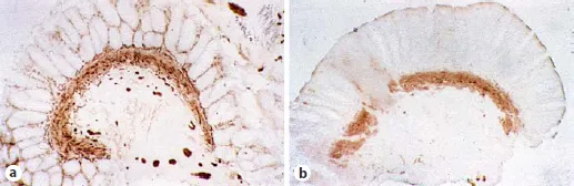
Fig. 2. a HD. Characteristic increase of AChE in parasympathetic nerve fibers of lamina propria mucosae and muscularis mucosae (2-week-old boy; compare with fig. 97). b Normal innervated rectum mucosa; 4-week-old boy. AChE staining. × 45.
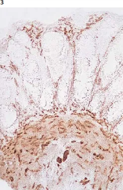
Fig. 3. Mucosa biopsy from the rectum (native cryostat section) with characteristic increase of AChE activity in parasympathetic nerve fibers in muscularis mucosae and lamina propria mucosae: HD. AChE reaction without counterstaining. × 90.
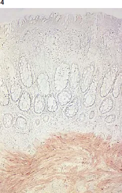
Fig. 4. AChE reaction of normal innervated rectum mucosa (native cryostat section). Only in muscularis mucosae are weakly stained nerve fibers to be observed. × 90.
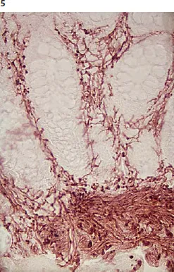
Fig. 5. Higher magnification of aganglionic mucosa biopsy section without counterstaining (7-month-old boy). × 180.
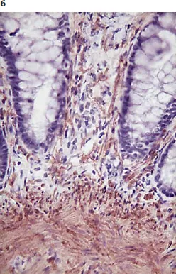
Fig. 6. Aganglionic rectum mucosa as in figure 5 with hemalum counterstaining. The identification of increased AChE activity in nerve fibers is much more difficult than in figure 5 without counterstaining. × 180.
In order not to miss a distal aganglionosis, the first biopsy may be taken from the rectoanal transition. Biopsies which are taken proximal from the anal ring may be taken in geometric distances (2, 4, and 8 cm above the anal ring). A seromuscular biopsy of the sigmoid allows no exclusion of an aganglionosis in the distal rectum [25, 26].
An enzyme histochemical AChE reaction in native rectal...
Table of contents
- Cover Page
- Front Matter
- A. Histopathological Diagnosis
- B. Different Colon Diseases with Chronic Constipation
- C. A Laboratory Guide to Histopathological Diagnosis of Intestinal Motility Disorders
- D. Preparation of Cryostat Sections from Biopsies and Colorectal Specimens
- E. Immunohistochemical Techniques of Paraffin Sections in the Diagnosis of Gut Dysmotility
- References
- Index
- Abbreviations
Frequently asked questions
Yes, you can cancel anytime from the Subscription tab in your account settings on the Perlego website. Your subscription will stay active until the end of your current billing period. Learn how to cancel your subscription
No, books cannot be downloaded as external files, such as PDFs, for use outside of Perlego. However, you can download books within the Perlego app for offline reading on mobile or tablet. Learn how to download books offline
Perlego offers two plans: Essential and Complete
- Essential is ideal for learners and professionals who enjoy exploring a wide range of subjects. Access the Essential Library with 800,000+ trusted titles and best-sellers across business, personal growth, and the humanities. Includes unlimited reading time and Standard Read Aloud voice.
- Complete: Perfect for advanced learners and researchers needing full, unrestricted access. Unlock 1.5M+ books across hundreds of subjects, including academic and specialized titles. The Complete Plan also includes advanced features like Premium Read Aloud and Research Assistant.
We are an online textbook subscription service, where you can get access to an entire online library for less than the price of a single book per month. With over 1.5 million books across 990+ topics, we’ve got you covered! Learn about our mission
Look out for the read-aloud symbol on your next book to see if you can listen to it. The read-aloud tool reads text aloud for you, highlighting the text as it is being read. You can pause it, speed it up and slow it down. Learn more about Read Aloud
Yes! You can use the Perlego app on both iOS and Android devices to read anytime, anywhere — even offline. Perfect for commutes or when you’re on the go.
Please note we cannot support devices running on iOS 13 and Android 7 or earlier. Learn more about using the app
Please note we cannot support devices running on iOS 13 and Android 7 or earlier. Learn more about using the app
Yes, you can access Histopathology of Chronic Constipation by W. A. Meier-Ruge,E. Bruder,W.A., Meier-Ruge,E., Bruder in PDF and/or ePUB format, as well as other popular books in Medicine & Gastroenterology & Hepatology. We have over 1.5 million books available in our catalogue for you to explore.